abot79-3

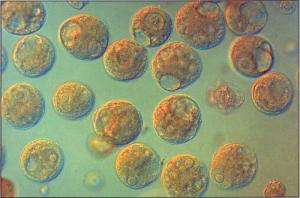
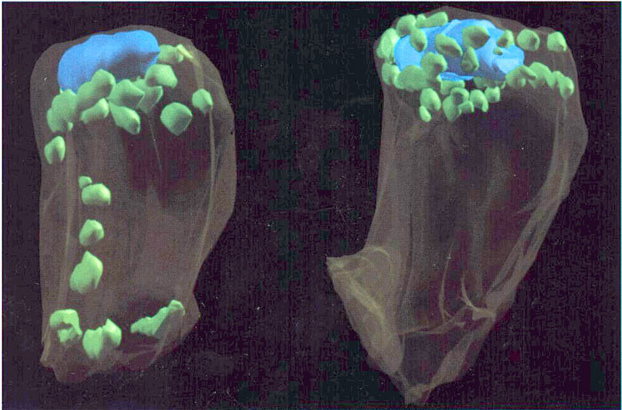
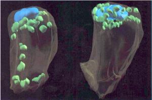
Microspores of Hemerocallis Fulva, isolated from anthers and converted to naked protoplasts by enzymatic treatment that removed the exine and entine (Nomarski interference contrast micography)
Article Title: Experimental Plant Reproductive Biology and Reproductive Cell Manipulation in Higher Plants: Now and the Future
Abstract: Experimental plant reproductive biology has recently emerged as a new form of experimental embryology characterized by the direct experimental isolation and manipulation of reproductive cells and protoplasts. This work is fostered by current multidisciplinary trends in the sciences and serves to deepen our knowledge about the control of reproductive processes by providing novel in vitro material. The techniques of experimental plant reproductive biology also show great potential in providing new means for biotechnology, leading to significant refinements in plant breeding in higher plants that may eventually permit direct reproductive cell engineering. Recent achievements by our research group in experimental manipulation and biological studies of pollen protoplasts, generative cells, sperm cells, and embryo sacs are reviewed, and some ideas about future developments in this new area are presented.
abot83-3
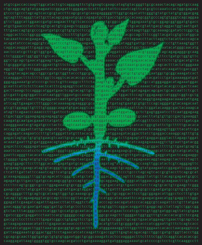
Computer-generated reconstruction from serial ultrathin sections showing the distribution of plastids within the egg cells of two genotypes of alfalfa. In the egg on the left, plastids (green) are positioned mostly below (toward the micropyle) the midtransverse region of the nucleus (blue) which results in the female plastids being largely sequested within the basal cell of the two-celled proembryo and not inherited. In the cell on the right, the plastids are perinuclear, thus, many female plastids become included in the apical cell of the two-celled proembryo and are inherited.
abot92-8
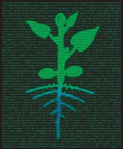
The classic schematic plant of J. von Sachs (Physiology of Plants. 1887. Clarendon Press, Oxford, UK) is superimposed onto repeats of the phytochrome DNA sequence to represent the plant body as an
abot95-01
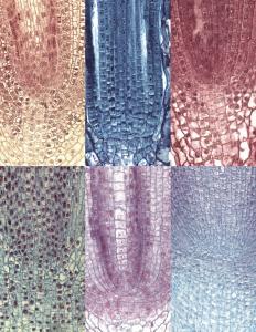
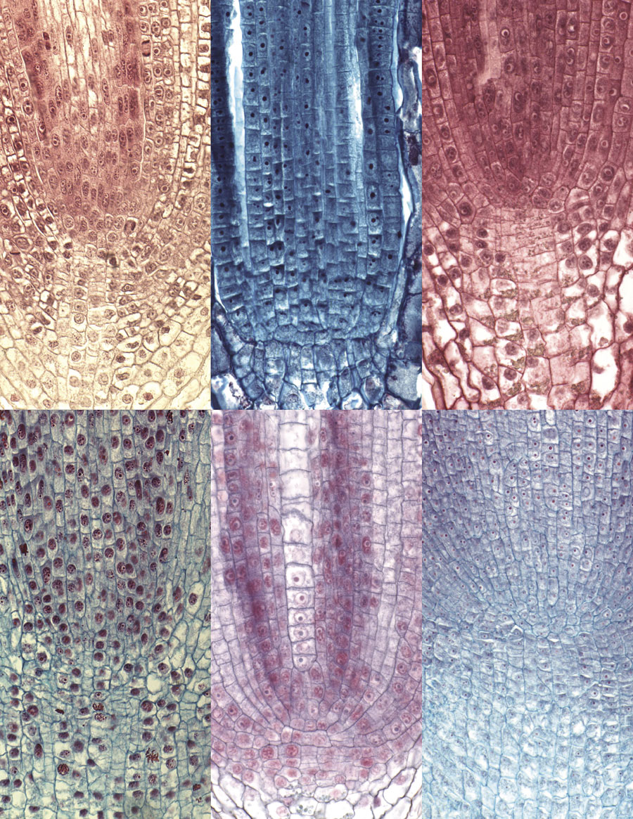
Light micrographs of median longitudinal sections of root apical meristems
(RAMs) from basal angiosperms, monocots, and eudicots are pictured, left to
right: top, Magnolia grandiflora, Cabomba caroliniana, and Ranunculus
muricatus; bottom, Clivia miniata, Commelina communis, and
Quercus rubra. This project, to characterize the arrangement of patterns
of cells in the organization of angiosperm RAMs, was conceived by Heimsch in
the early 1950s; the specimens in this montage of root tips were produced from
1956 until 2003 and were photographed with brightfield illumination on a Zeiss
Axiophot microscope (University of Waterloo).
For further details, see: Heimsch and Seago, Organization of the root
apical meristem in angiosperms, American Journal of Botany,
Volume 95, Issue 1, pages 1-26, http://www.amjbot.org/cgi/content/short/95/1/1.