14-001h

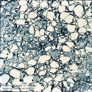
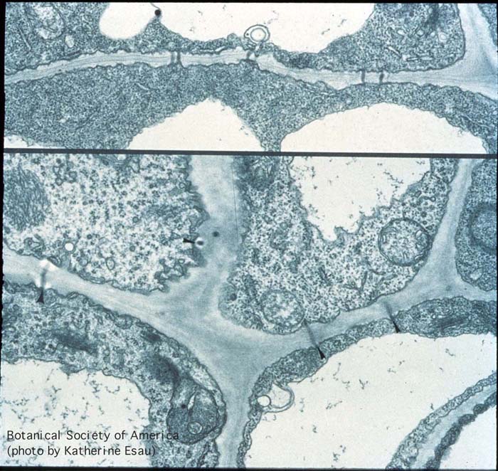
Young parenchyma tissue cut parallel with the epidermis of Euphorbia pulcherrima (poinsettia). Plasmodesmata present in cell walls remain united between cells during intercellular spaces development.
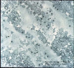
14-002h
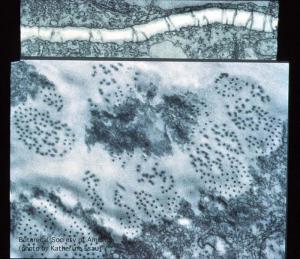
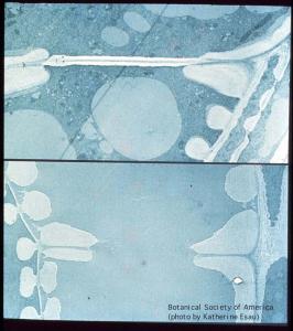
Parenchyma cells in phloem of Cucurbita maxima leaf. In one view, plasmodesmata are shown in longitudinal views, and in transverse views in primary pit fields.
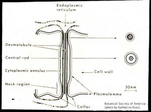
14-003h
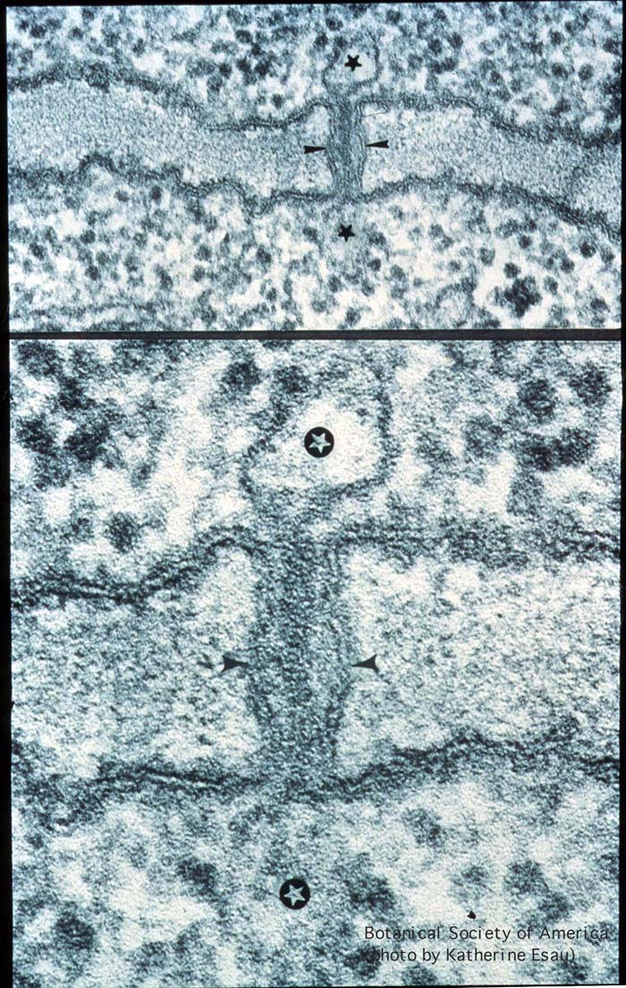
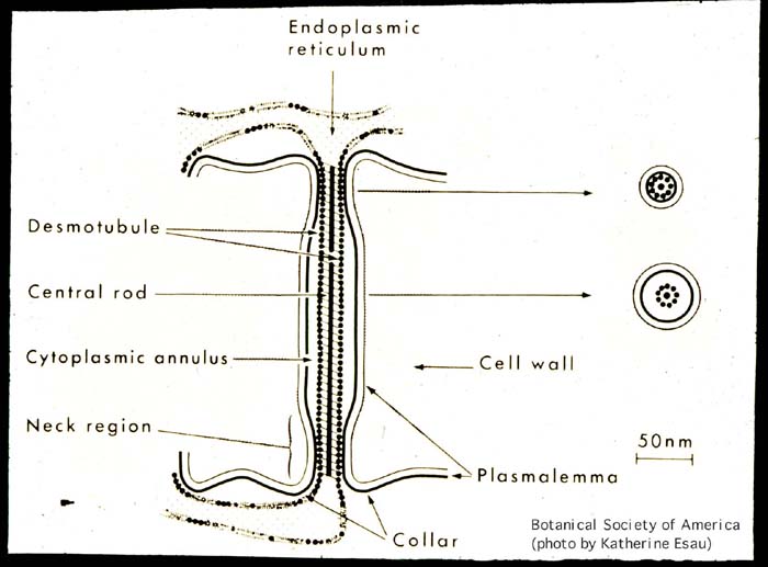
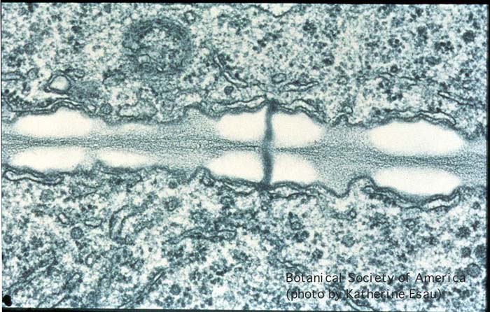
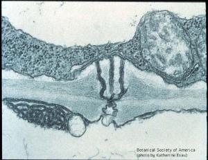
Plasmodesmatal structure in longitudinal section of Phaseolus vulgaris root tip. Cell walls are lined with plasmadesmata between contiguous cells. Demotubule extends to the lumina of ER cisternae.
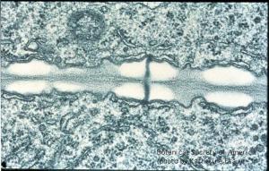
14-004h
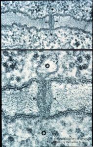
Constrictions at both ends of plasmodesma presumably have valve-like function.
14-005h

Plasmodesmata originating during cell division. Mitosis associated with accumulation of vesicles in the equatorial plane. This is first stage in cell-wall formation of Nicotiana tabacum mesophyll.
14-006h
Mitosis in longitudinal divisions of elongated cells. Earlier stage in Nicotiana tabacum; later stage (more extended cell plate) in Sonchus oleraceus.
14-007h
Late telophase (nuclei not shown). Microtubules assemble into a phragmoplast centrifugally around the cell plate toward the lateral mother-cell walls (petiole of Echium judaeum).
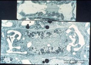
14-008h
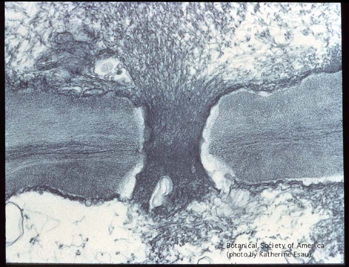
Longitudinal and partial surface views of growing cell plates showing involvement of ER in formation of plasmodesmata. ER tubules may be included in openings in the cell plate. Phaseolus vulgaris root tip.
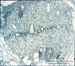
14-009h
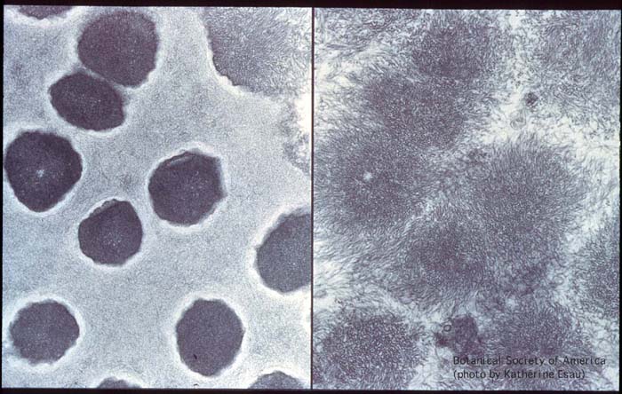
Cell plate has become the cell wall with fully formed plasmodesmata. Incomplete section made parallel with the surface of the cell wall. Phaseolus vulgaris root tip.
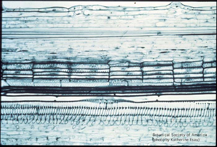
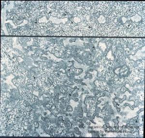
14-010h
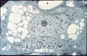
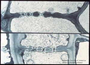
Young sieve element and companion cell, the latter narrow, with a narrow nucleus. Cell wall between the two cells and plasmodesmata in future sieve plates. (Nicotiana tabacum root tip)
14-011h
Two stages in sieve plate development: plasmodesmal stage and plasmodesmata partly lined with callose. (Nicotiana tabacum root tip)
14-012h

Young wall with plasmodesmata (top); sieve plate elements (left) and companion cells (right). Early stage of callose accumulation visible at ends of a plasmodesma, near differentiating pore site. (Echium angustifolium and E. sabulicola petioles)
14-013h
Further development of a pore site in longitudinal view. Callose nearly fused at middle lamella, plasmalemma and ER cisternae cover the callose, plasmodesma extends from ER to ER through future pore. (Echium angustifolium petiole)
14-014h
Continuous callose and plasmalemma in former plasmodesmal canal (above). Most callose was removed and open pores partly occluded with P-protein. (Echium angustifolium petioles)
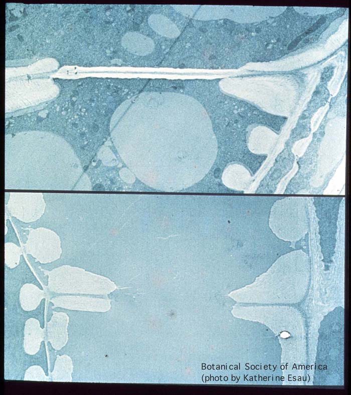
14-015h
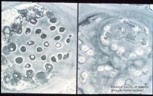
Left, plugs of callose, penetrated by plasmodesmata, fill the pores. Right, pores open, lined with thin layer of callose, and most are occluded with P-protein, some with starch grains. (Echium wildpreti petioles)
14-016h

P-protein plug in pore in longitudinal section. Compaction of P-protein within pore especially through middle lamella region. Echium judaeum petiole
14-017h
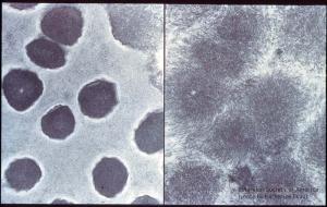
Cross sections through pores filled with P-protein and through masses of loose P-protein located at some distance from the sieve plate. Echium plantagineum petioles.
14-018h
P-protein digestion with pepsin (left); control (right). Compacted P-protein within the sieve plate pore became digested. (Echium judaeum petiole)
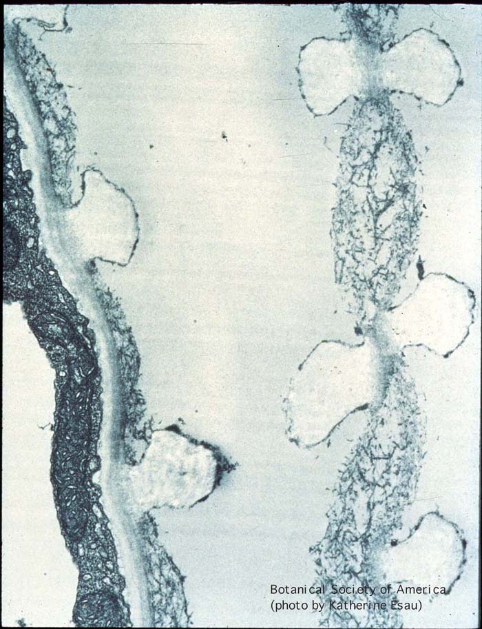
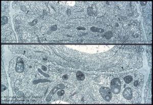
14-019h
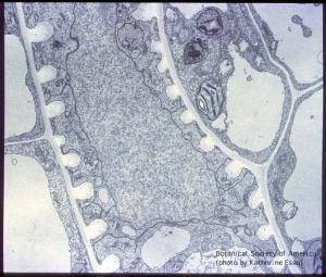
Longitudinal section of cell wall including a plasmodesma connecting a sieve element (below) and branched plasmodesma located on the companion cell side of the wall. (Echium rosulatum petiole)
14-020h
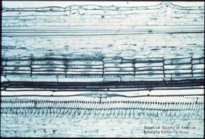
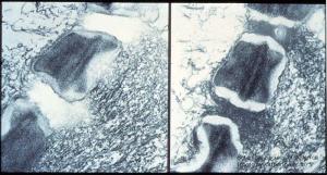
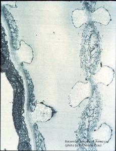
Primary xylem of Phaseolus vulgaris stem, with ring-like and helical secondary walls (close to bottom of slide). Secondary wall surrounds perforations at the two ends of an individual vessel member. Perforations are formed by localized removal of the primary wall.
14-021h
Removal of vessel end wall with middle part consisting of primary wall material. Around the end wall margin, secondary wall material protects unthickened primary wall, which becomes hydrolyzed and disappear. This stage is shown below: the rim now surrounds a perforation. (Phaseolus vulgaris stem)
14-022h
Unperforated vessel wall parts also undergo a change; primary wall not covered by secondary wall layers undergoes partial hydrolysis. Beta vulgaris leaf.
14-023h
Primary wall between contiguous vessel members (right) is partially hydrolyzed between secondary thickening gyres. Noncellulosic wall components removed, cellulose forms a loose network. Hydrolysis affects primary wall adjacent to vessel member. (Capsella bursa pastoris leaf)
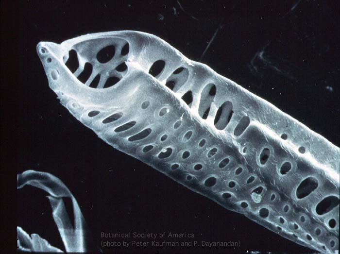
14-024h
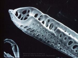
Scanning electron microscograph of vessel member from Pelargonium leaf with perforations and pits. Micrograph, courtesy of Professor Peter B. Kaufman and Dr. P Dayanandan, Dept. of Botany, Univ. of Michigan, Ann Arbor, MI.
26-005
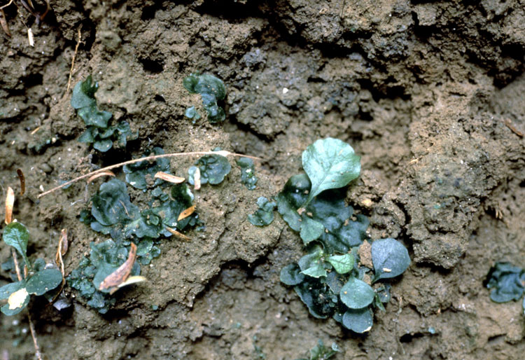
Ophioglossum fertile spike, l.s.
26-009
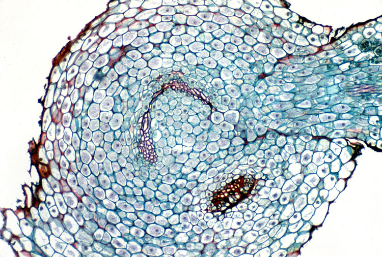
Marattia synangium x.s.
26-012
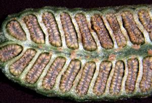
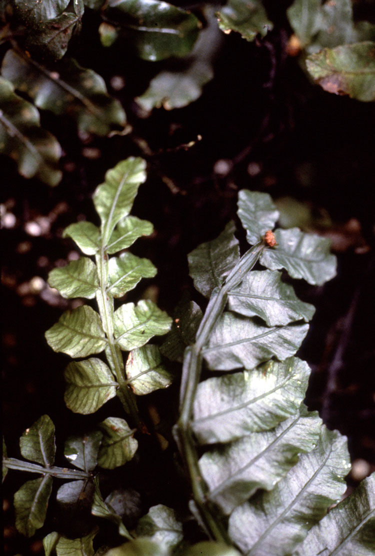
Danaea wendlandii fertile pinna