FALL 2023 VOLUME 69 NUMBER 3
PLANT SCIENCE
BULLETIN
A PUBLICATION OF
THE BOTANICAL SOCIETY OF AMERICA
Art
in the
Botanical
Sciences
Applications in Plant
Sciences
Celebrates Its 10th
Anniversary... p. 252
Botanical Society of America’s Award
Winners... p. 244
Fall 2023 Volume 69 Number 3
FROM the EDITOR
Greetings,
I am thrilled to share this very special issue of PSB that focuses on the many connections
between science and art. This issue has been brought to you by an impressive team of guest
editors: Patricia Chan, Rosemary Glos, Ashley Hamersma, Kasey Pham, and Nicolette
Sipperly. I want to thank this team for their creative visioning and hard work putting this
issue together. I also want to thank the talented authors and creators who have shared their
work with us. There was such a positive response to the call for articles on this topic that we
anticipate continuing this subject in our Spring and Summer issues.
In this issue, you will also find many of our regular PSB sections, as well as news and awards
from Botany 2023 and profiles of our new Distinguished Fellows.
Sincerely,

PSB 69 (3) 2023
159
159
TABLE OF CONTENTS
SPECIAL SECTION: ARTS IN THE BOTANICAL SCIENCES: PAST, PRESENT,
AND FUTURE................................................................................................................................
161
Weaving Together Culture and Ecology to Express My Identity as a Scientist
(by Clarissa Rodriguez)......................................................................................................................... 162
Art in the Herbarium? (by Maura Flannery)....................................................................................... 165
The Significance of Illustrations as Nomenclatural Types in Botany: “Iconotypes”
at the Natural History Museum Vienna and the Importance of Color Systems,
such as Those Utilized by Ferdinand Bauer [1760–1826]
(by Tanja M. Schuster et al.)............................................................................................................... 170
Celebrating the Launch of the UTEP Virtual Herbarium by Highlighting
Contemporary and Historical Art and Science of the El Paso Region
(Mingna V. Zhuang et al.)..................................................................................................................... 176
Celebrating Plant Diversity through Art (by Alice V. Pierce et al.) ........................................ 179
Brazilian Botanists Flirting with Arts: Valuing the Multicultural Heritage
(by Lucas C. Marinho et al.) ................................................................................................................ 182
Integrating Botany, History, Culture, and Contemporary Art in a Botanical Garden
Museum (by Nezka Pfeifer)............................................................................................................... 186
Science, Art, and the Allure of Photographs (by Benjamin-Goulet-Scott
and Jacob Suissa).................................................................................................................................... 189
Filling Out PhyloPic: Call for Adoption by Plant Scientists (by Mason C. McNair
and T. Michael Keesey)......................................................................................................................... 193
Can the Collaboration of Science and Art Broaden Our Understanding of Nature?
(by Paul J. CaraDonna and Mark Dorf) ........................................................................................ 198
The Integration of Botanical Science, Art, and Agency (by Maria Park et al.)................ 202
“Art of Horticulture” Course Cultivates Creativity (by Emily Detrick
and Craig Cramer..................................................................................................................................... 206
Reaching Across Audiences: Connecting to and Communicating Botanical Concepts
Through Art (by Janette L. Davidson et al.) .............................................................................. 209
Reconnecting Science with the Visual Arts: Teaching the Art of Biology
(by Lynne Gildensoph) .......................................................................................................................... 216
Nature Journaling: Sharing Perspectives Between Art and Science
(by Corinn Rutkoski et al.).................................................................................................................... 219
Plants as a Case for Creative Collaboration: Designing the Interactive Art-Science
Exhibition Meaningful Beauty (by Christopher Ault et al.) ................................................ 221
Understanding Plants Through Imagery: Functional Traits, Cuteness, and
Narrative (by F. Curtis Lubbe) .......................................................................................................... 227
Why Are You Doing That? Unexpected Varaition in Floral Traits of
Blue Cardinal Flower (by Gavin Hossack).................................................................................. 232
Wild Growth Paintings (by Daniel Philosoph)................................................................................... 237
Art + Botany: Making a Difference (by Kathleen Marie Garness) ......................................... 239

PLANT SCIENCE BULLETIN: FALL 2023
..............................................................................................244
SOCIETY NEWS
Botanical Society of America’s Award Winners (Part 2) ........................................................................244
Publications Corner..................................................................................................................................................... 252
Botany 2023At A Glance......................................................................................................................................... 254
MEMBERSHIP NEWS
BSA Spotlight Series.................................................................................................................................................. 257
BSA Professional Highlight Series...................................................................................................................... 258
New AJ Harris Graduate Student Award........................................................................................................ 259
BSA Student Chapter Updates..............................................................................................................................259
It's Renewal Season .....................................................................................................................................................260
PSB
Print Subscription Change
............................................................................................................................
260
Year-End Giving.............................................................................................................................................................260
2023 BSA Gift Membership Drive .......................................................................................................................261
New BSA Ad Hoc Committee................................................................................................................................261
SCIENCE EDUCATION
Updating BSA’s State-by-State Botanical Resource Pages Please Help!.................................... 264
PlantingScience Updates......................................................................................................................................... 264
Meet Our F2 Fellows .................................................................................................................................................. 265
Read About Digging Deeper.................................................................................................................................. 266
STUDENT SECTION
Botany 2023 Review................................................................................................................................................... 268
IN MEMORIAM
Dr. Joel Fry (1957-2023)....................................................................................................................................... 270
ANNOUNCEMENTS
Dr. John Kiss Featured on The Space Show............................................................................................. 273
BOOK REVIEWS.................................................................................................................................................
274
S
tart Planning for
See logo description on inside back cover

161
From the P SB Special I ssue on Art in the Botanical Sciences
SPECIAL SECTION
Art in the Botanical Sciences:
Past, Present, and Future
As you may have guessed from the cover, this is no ordinary issue of the PSB! You are reading the first
in a multiple-issue anthology dedicated to art and the botanical sciences. We, your guest editors, are a
group of graduate students from four universities who are passionate about the intersections of botany,
art, and personal expression. This anthology grew out of our first workshop on botanical art at Botany
2022 in Anchorage, AK. As artist-scientists, we have worked to create spaces where our colleagues can
discuss their complex, and sometimes challenging, experiences of bridging the gap between disci-
plines. The PSB editorial team invited us to expand on the ideas shared in our workshop via a special
issue. We accepted with enthusiasm and set to work soliciting abstracts for an issue that celebrated
the many ways that people integrate art and botany. We received so many exciting proposals that the
resulting pieces will be published in three(!) consecutive issues of the PSB.
In this issue, you will learn about a variety of artist-scientist collaborations, integrations of art and
science in museums, scientists’ and artists’ accounts of what drives their curiosity and exploration, and
the role of art in teaching and herbarium curation.
In preparing this issue, we learned that “anthology” comes from the Greek anthologéō, or “I gather
flowers.” How fitting for a collection of works that celebrate the beauty and wonder of the botanical
world. We hope the pieces featured in this anthology inspire you to envision how creativity can enrich
our lives as scientists, artists, and educators.
Enjoy!
The SciArt Collective
Nicolette Sipperly, Stony Brook University
Rosemary Glos, University of Michigan
Kasey Pham, University of Florida Patricia Chan, University of Wisconsin-Madison
Ashley Hamersma, University of Florida

162
From the P SB Special I ssue on Art in the Botanical Sciences
ABSTRACT
To reach broader audiences, science
communication must move towards imaginative
and unconventional methods of conveying
research and knowledge. I use embroidery to
weave together my personal, scientific, and cultural
experiences and share them with not only other
scientists but general audiences and my family.
I grew up helping my grandparents forage for
medicinal plants, weed their crop fields, and herd
our family cattle every summer in the grasslands
of Mexico. Our daily work required knowledge
of when and where plants grow, when to harvest,
and when to rotate cattle. Despite my exposure to
botany and ecology at an early age, I never linked
my family’s cultural knowledge to these fields of
science. The more I researched about my Mexican
heritage, the more proof I found of various ways
to transfer knowledge via cultural practices.
Traditional embroidery has long been a way
for people to record the plants and animals that
Weaving Together Culture and Ecology to
Express My Identity as a Scientist
were present on the land, as well as an important
way to share important stories and lessons. As a
scientist, I integrate my culture into my work by
using embroidery to communicate my research,
study species, and express my identity through
art. This form of transferring knowledge also
helps promote cultural diversity and inclusivity
in science. Given that information is conveyed
mainly through visualizations rather than text
via embroidery and other art forms, SciArt also
removes language barriers that may prevent
individuals from engaging with science. Although
I use embroidery here as an example of expressing
my identity as a scientist, I encourage others to
find ways to express their science using other
forms of media (e.g., sculpture, dance, song) that
provide creative avenues for passing knowledge
from one generation to the next.
I was fortunate to spend my childhood summers
in the semi-arid grasslands of Zacatecas, Mexico
with my grandparents. Surrounded by a diverse
range of flora and stunning exposed cliffs, I
was captivated. My grandparents taught me the
importance of being land stewards, showing me
how to rotate cattle to ensure plant regrowth
the following year and how to identify and use
plants with ethnobotanical properties. These
were all lessons that had been passed down
orally for generations in my family. Although
I was not consciously aware of it at the time,
these experiences were integral to developing
my scientific worldview, but I would not begin to
link my family’s cultural knowledge to scientific
concepts in ecology until college. This realization
Clarissa Rodriguez
Plant Biology Ph.D. Candidate,
University of California-Riverside

163
allowed me to self-reflect on my identity and
explore other forms of transferring knowledge in
Mexican culture that I may have overlooked. I was
captivated by the use of artisan’s hand embroidery
to convey stories and even record flora and
fauna that were present on landscapes through
beautiful images created on textiles using colorful
threads. Today, I am continuing this tradition by
using embroidery to communicate my science
with broader audiences, express my identity as a
Mexican-American scientist, and weave together
my scientific training and heritage in hopes
of encouraging the representation of cultural
diversity and knowledge in science.
As a first-generation Mexican-American, I wanted
to fit in with my peers, and sometimes fitting in
came at the cost of suppressing my own Mexican
heritage. My mother started to teach me how to
embroider when I was 10, showing me the various
stitching techniques my great-grandmother had
taught her. As a child, I was eager to learn how
to use stitches to display intricate flowers, birds,
and mammals. But as I entered my teenage years,
I spent less time practicing my embroidery until I
stopped altogether to make time for other hobbies
that I could share with my American friends. I had
also stopped making my annual trips to Mexico
due to heavy cartel violence that was rampant at
the time. This change meant that I could no longer
engage in ranching, farming, or family gatherings
in Mexico—activities that had always been a
central part of my life. Consequently, this led to
an identity crisis where I found myself identifying
more with my American companions and losing
the firm grip I had on my Mexican roots.
During my college years, I wasn’t entirely sure
about my cultural identity, but I knew for certain
that I wanted to become a scientist to solve the
environmental concerns plaguing our world.
My understanding of science was limited to the
Western perspective, where trained scientists
collected data and validated it through peer review.
However, it wasn’t until I took an undergraduate
ecology course that I realized how much my
grandparents’ traditional practices were rooted in
the fields of community and restoration ecology. It
was foolish of me to not recognize earlier that their
knowledge of rangelands and natural resource
management was science. Their ecological
knowledge was accumulated over generations of
trial and error, and this realization encouraged
me to reflect on my identities. In turn, I found
a creative outlet through SciArt embroidery to
express my cultural ties and share science.
My first attempt at merging science and art via
embroidery was unsuccessful. I had forgotten
how technical the work of embroidery was, and
my work was riddled with crooked stitches and
tension issues throughout. Although it wasn’t
pretty, I wanted to show my grandma that I had
finally found a way to share my love for science
while paying homage to my Mexican roots. After
ten years of not seeing my family in Mexico, I
made the journey to reconnect, share my work,
and seek advice on how to fix my technique. My
grandma and my aunts gave me pointers on how
to improve my technique, but also expressed joy
that “the young generation is keeping our tradition
alive.” My mother was especially proud of me for
embracing my Mexican heritage within academic
spaces, because she had witnessed my struggles
with identity for years. This trip inspired me
to design a series of hoops to convey important
concepts and processes within the field of plant
ecology.
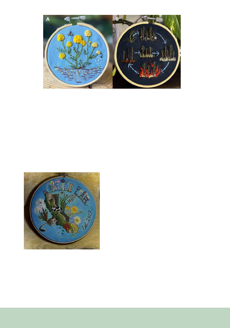
Given my scientific interests, plant invasions are
a common theme in my SciArt. For instance,
stinknet, a focal species of my dissertation, is an
invasive plant that is difficult to manage due to its
prolific seed production and persistent soil seed
bank (Figure 1A). Likewise, I created a hoop that
shows how the dispersal of introduced grasses
into a community can establish an invasive grass-
fire feedback loop by increasing biomass and fuel
continuity while suppressing native plant recovery
(Figure 1B).
A large reason why I feel comfortable expressing
myself within academic spaces is due to my
advisor’s support, Dr. Larios, who is a fellow first-
164
Figure 1. Embroidery hoops showing (A) stinknet (Oncosiphon pilulifer) accumulating seeds in the seed bank, and
included a harvester ant (Pogonomyrmex rugosus) crawling along the soil surface to depict a collaborative study in-
vestigating the role of harvester ants in dispersing invasive plants, and (B) invasive grass-fire cycle that establishes once
invasive grasses disperse into a community, which creates a litter layer that adds fuel and connectivity that carries fire
easily and creates a feedback loop to maintain invader dominance while suppressing native recovery.
generation Mexican-American, and an incredible
scientist. To show my appreciation for her
dedication to creating an inclusive workspace, I
designed and embroidered a hoop to represent the
various research topics (e.g., trophic interactions,
coexistence and land management) she studies
Figure 2. A 12-inch embroidery hoop I made for my
PhD advisor, Dr. Larios, to showcase the wide scope of
her community and restoration ecology research pro-
gram in California, which was established in 2017.
throughout California (Figure 2). One of my
favorite things about embroidery is the removal
of a language barrier. One can stare at a hoop
and recognize the heterogenous landscape of
California, along with the biodiversity of plant
species, herbivores (e.g., cows, kangaroo rats)
and fire on the landscape hinting at ecological
processes.
SciArt is a powerful tool for self-expression
and knowledge transfer that can reach broader
audiences and increase inclusivity in science. For
many individuals who come from historically
underrepresented groups within the fields of
STEM, this is a way for us to help integrate forms
of cultural knowledge and dissemination into
a Western-dominated society. Peer-reviewed
journals are already recognizing the power of
art to convey information by requiring graphical
abstracts (i.e., visual representations of the article’s
main findings). Although I use embroidery here, I
encourage others to find their own artistic outlet
to promote their science and help them share
their story. I urge my colleagues to try out visual
and performing arts to discover their preferred
creative outlet. Above all, do not hesitate to
take the first steps toward integrating art with
botanical sciences, whether that means making
the initial brush stroke, threading the first needle,
or collaborating with an artist.
165
From the P SB Special I ssue on Art in the Botanical Sciences
In the 1980s, only one species of the genus Anisotes
was known in Madagascar. However, a specimen
on loan to the California Academy of Sciences
(CAS) from the National Museum of Natural
History in Paris seemed to be from the same genus
but with distinctive leaf and flower shape. Daniel
Thomas of the CAS wanted to know more, so he
asked botanical illustrator Erin Hunt to draw the
plant from the specimen. The result was different
from what he expected, but Hunt thought she
had captured the form correctly. When CAS
botanists went to Madagascar with the drawing
in hand, they found the plant, identifiable from
the drawing. Hunt had been right (Daniel et al.,
2007).
Such stories are rather common in botany.
After the illustrator Patricia Fawcett at Fairchild
Botanical Garden drew a flower from life with
Maura Flannery
Doctor of Philosophy, St. John’s University,
Department of Computer Science,
Mathematics and Science, New York, NY
Art in the Herbarium?
too many floral parts to fit the taxon description,
botanists realized intra-species variation required
amending the description (Stevenson and
Stevenson 2014). Conceptualizing a given species
can involve several different kinds of input ideally
including fresh and preserved material, but also
hand-drawn illustrations. The emergence of
modern botany in the 16th century was based on
this assumption, which required the rejection of
classical authorities’ deep distrust of hand-drawn
illustrations (Reeds, 1976). It is not surprising
that Luca Ghini (d. 1556), the director of the first
botanic garden in Pisa, was a very early proponent
of herbaria and field trips as well as a collector of
drawings and printed artwork, some from the first
printed herbals with good illustrations (Findlen,
2017). It quickly became common for botanists
to trade or borrow both specimens and images,
as they moved away from reliance on classical
authorities who distrusted plant drawings.
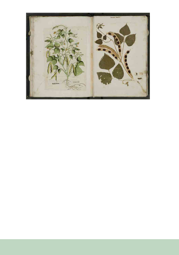
The Swiss naturalist Felix Platter (1536-1614)
arranged his bound collection so that in many
cases he would have one or more images on the
left-hand page, and a specimen of the same species
on the right (Figure 1; Benkert, 2016). On the
other hand, the Italian naturalist Ulisse Aldrovandi
(1522-1605) stored his specimens and watercolors
separately. Some early modern botanists used
other visual technologies including nature prints
and rubbings to get information about plants into
a portable reference format. There were even
some who painted in missing parts of a specimen.
Because botanical terminology was in its infancy,
visual representations could differentiate among
similar species in ways that written descriptions
166
could not. Those interested in plants preferred
to receive drawings rather than text. Some argue
that it was the illustrations that pushed botanists
to write descriptions of equal clarity, and that
the artists of the age were influential in causing
botanists to observe more closely (Smith, 2003).
The relationship between drawing and knowing
has become a topic of interest among historians
of art and science. The astronomer Omar Nasim
(2013) describes how repeated drawing of nebulae
in the 19th century clarified the concept of an
astronomical phenomenon that, as its name
suggests, was blurry. As more observations were
made, as drawings were repeated night after night,
these hazy structures become more familiar to the
observer’s eye, mind, and hand. Drawings were
tools in the process of discovery and knowledge
stabilization. In drawing, the hand slows down
observation, allowing the mind to synthesize.
Barbara Wittmann (2013) reports on a scientific
illustrator’s experiences working on a new fish
species where getting the drawings correct led him
to discover novel anatomical features. Botanists
who choose to do at least their own preliminary
drawings have similar experiences. As they draw
plant structures repeatedly, often from different
perspectives or from different dissections, they
find themselves learning more and having to
correct their drawings and written descriptions
along the way.
If a professional illustrator is doing artwork, there
is a back-and-forth, with the botanist making
corrections. This is what Lorraine Daston and Peter
Galison (2007) term “four-eyed sight,” producing
a more informative image than one pair of eyes
could. Clarity and discovery arise from this give-
and-take. A drawing helps both the botanist and
the audience to understand more about the plant.
The zoologist and illustrator Jonathan Kingdon
(2011) appeals to research on the neurophysiology
of sight to explain why pen and ink drawings and
prints are particularly useful. The human brain
processes signals from the eyes by detecting edges
and accentuating them. This means that black-
and-white illustrations communicate information
especially effectively because they are in tune with
the brain’s visual processing system.
Figure 1. From Felix Platter’s herbarium (volume 7, pp. 86-87). Hand-col-
ored woodcut image of common bean (Phaseolus vulgaris) from the Ger-
man edition of Leonhard Fuchs’s 1542 herbal, facing specimen of the same
species. Courtesy of Bern City Library.

167
As more and more new plants were discovered
around the world beginning in the 1500s, botanists
were challenged with naming and describing
them. There was a fervor among many to publish
new species as soon as possible, often based only
on specimens or drawings sent to them. Even
Carl Linnaeus described many species solely on
the basis of watercolors or printed black-and-
white illustrations. When his species descriptions
were later typified, these images became
lectotypes since they were the only materials he
had examined. This was true for other botanists
as well, although now the practice is only allowed
in defined cases in the International Code of
Botanical Nomenclature for Algae, Fungi, and Plants
(Turland and Wiersema, 2018).
Pragmatically, drawings of plants also serve as a
back-up to specimens and descriptions. The plants
of New Spain that were collected in the late 18th
century by the Sessé and Mociño Expedition
reached Spain safely, but their analysis and
publication was disrupted by Martin de Sessé’s
death and the Napoleonic Wars. José Mociño
fled to France, taking with him some written
descriptions and nearly 2000 illustrations made
during the expedition. In Montpellier, he shared
this hoard of information on undescribed species
with Augustin Pyramus de Candolle, who was
writing a comprehensive work on the world’s
plants. De Candolle regarded these watercolors
and sketches as so valuable that he had them hastily
copied by a large team of artists before Mociño
returned to Barcelona. After Mociño’s death there,
the originals were lost until 1979! Consequently,
for more than 150 years, de Candolle’s annotated
copies became the primary source of information
about hundreds of New World species (McVaugh,
1998).
In the 18
th
and 19
th
centuries, British colonial
administrators in India created botanical gardens
as a way to gather and observe many species to find
those that could be sources of food, medicines,
and useful items such as textiles and timber
wood. Meanwhile, botanists collected thousands
of specimens in support of this effort. It became
standard practice to have Indian artists paint
watercolors of many of these plants. This was not
just to have a record of a plant’s color and form,
but also to ensure that there was any record at all.
In tropical climates, conditions sometimes drove
collectors to rely on nature prints and drawings. It
was difficult to preserve specimens in hot humid
areas and to prevent insect and fungal damage.
In the 20
th
century there were botanists like Oakes
Ames at Harvard who included ink drawings and
watercolor sketches by his artist wife Blanche Ames
on his orchid specimens. This approach continues
as drawings of flower dissections and microscope
enlargements of structures are added to sheets by
botanists and artists. Because of the importance
of having different kinds of information about a
plant available at the same time, such practices
persist, but there has been a change in storage
practices. In the past, illustrations of a species
were often stored in the same folder as specimens
of that species. This was handy for botanists but
perhaps not so good for the art, which could be
damaged by substances leaching from the plants.
With the start of digitization projects, when
folders that hadn’t received attention for years
were examined, it became common to remove the
illustrations, particularly if they weren’t physically
attached to the specimens. They are now usually
stored separately, often in the library or archives
affiliated with the herbarium.
The Royal Botanic Garden Edinburgh (RGBE)
has a large number of Indian plant specimens,
many collected by physicians who had trained
in Edinburgh and later served Britain’s massive
colonial enterprise in India. Henry Noltie, then
a curator at the RBGE herbarium, spent months
going through folders, removing the images created
by Indian artists and successfully re-sorting them
from taxonomic order into separate collections,
many donated by one person or organization
and in some cases created by a single Indian

168
artist or by a small group. Noltie was often able
to identify the artists by name, but others remain
anonymous. Through this work, Noltie learned
a great deal about the collections and the people
related to them, producing several books on these
works (Noltie, 2002, 2017). The art is now stored
in the RBGE archives, along with correspondence
and other papers from those responsible for the
specimen and art collections. The Royal Botanic
Gardens Kew and many other institutions have
done the same.
This policy makes the illustrations more available
to those interested in the historical and artistic
significance of these works and keeps them
together as collections so they can be compared
in terms of style. In other words, the images are
now treated more as works of art than of science,
perhaps to the detriment of science. What might
be good for the botanical art and for the history
of art and botany, might not be the best solution
for botanical inquiry. Past collectors of Indian
botanical art were often as interested in them as
works of art as of science, and today this is true
for many who collect botanical art. However, both
the art and specimens were created as scientific
documents, whether or not they had any obvious
aesthetic appeal. The botanist Peter Crane (2013)
considers botanical illustration as important
today as it was hundreds of years ago, but notes
economics has changed the picture. Today, color
images are rare in taxonomic treatments of new
species, and even pen-and-ink illustrations are
less prevalent, even though, for all the reasons
discussed earlier, illustrations can provide vital
information to botanists.
The works described here were created as adjuncts
to specimens, living and preserved, and they should
be able to continue to function this way. Opening
up a species folder and finding illustrations can
provide a botanist with information on color and
form less likely to be documented in the specimen
itself, thus making it easier to create a full mental
image of the plant. If the illustrations are stored
elsewhere, this richness is lacking. A trip to the
archives would be necessary to see the relevant
drawings. Since some of the art collections have
been digitized, and herbaria are continuing to
digitize their collections, it should be possible to
link a specimen to one or more illustrations of that
specimen as one facet of the extended specimen
concept.
Implementing such an idea is hardly trivial.
The databases used in science and those for art
and archival materials are often quite different.
There are moves toward standardization across
platforms, but implementation will not be easy.
The work of developing what is called FAIR
data—Findable, Accessible, Interoperable, and
Reusable—is substantial (Manzano and Julier,
2021). The colonial origin of a good deal of both
types of collections needs to be acknowledged as
well. However, my focus here is on the value of
linking art to science, an important component
since the 16th century. It is as important today
not only for what botanists can learn about
plants but for the aesthetic lift that comes with
examining these works, even if the goal is to seek
out taxonomic information. The botanist Richard
Mabey’s (2015) comment is relevant here: “The
quintessence of a plant can only ever be a fantastic
goal, something to travel towards but never reach”
(p. 27). That’s why we need to dig into—and
preserve—different types of information: living
plants, specimens, art, and photography, which
should not be forgotten but is outside the scope
of this article.
REFERENCES
Benkert, D. 2016. The ‘Hortus Siccus’ as a focal point:
Knowledge, environment, and image in Felix Platter’s and
Caspar Bauhin’s herbaria. In S. Burghartz, L. Burkart, and
C. Göttler (Eds.), Sites of Mediation, 211–239. Brill, Leiden.
Crane, P. 2013. Gingko: The Tree that Time Forgot. New Ha-
ven, CT: Yale University Press.
Daniel, T. F., B. A. V. Mbola, F. Almeda, and P. B. Phillipson.
2007. Anisotes (Acanthaceae) in Madagascar. Proceedings of
the California Academy of Sciences 58: 121–131.

169
Daston, L., and P. Galison. 2007. Objectivity. Zone, New
York.
Findlen, P. 2017. The death of a naturalist: Knowledge and
Community in Late Renaissance Italy. In G. Manning, C.
Klestinec (Eds.), Professors, Physicians and Practices in the
History of Medicine, 127–167. Springer, New York.
Kingdon, J. 2011. In the eye of the beholder. In M. R. Can-
field (Ed.), Field Notes on Science and Nature, 129–160. Har-
vard University Press, Cambridge, MA.
Mabey, R. 2015. The Cabaret of Plants: Forty Thousand Years
of Plant Life and the Human Imagination. Norton, New York.
Manzano, S., and A. C. M. Julier. 2021. How FAIR are plant
sciences in the twenty-first century? The pressing need for
reproducibility in plant ecology and evolution. Proceedings of
the Royal Society B: Biological Sciences 288: 20202597.
McVaugh, R. 1998. Historical introduction. Torner Collection
of Sessé and Mociño Biological Illustrations, Hunt Institute for
Botanical Documentation. Website: https://www.huntbotani-
cal.org/art/show.php?10.
Nasim, O. W. 2013. Observing by Hand: Sketching the Nebu-
lae in the Nineteenth Century. University of Chicago Press,
Chicago.
Noltie, H. J. 2002. The Dapuri Drawings: Alexander Gibson
and the Bombay Botanic Gardens. Antique Collectors’ Club,
Edinburgh.
Noltie, H. J. 2017. Botanical Art from India: The Royal Bo-
tanic Garden Edinburgh Collection. Royal Botanic Garden
Edinburgh, Edinburgh.
Reeds, K. M. 1976. Renaissance humanism and botany. An-
nals of Science 33: 519-542.
Smith, P. H. 2003. The Body of the Artisan: Art and Experi-
ence in the Scientific Revolution. University of Chicago Press,
Chicago.
Stevenson, J. W., and D. Wm. Stevenson. 2014. The nuts and
bolts of doing the Flora of the Bahama Archipelago: How Don
Correll worked. The Botanical Review 80: 135–147.
Turland, N. J., and J. H. Wiersema (Eds.). 2018. International
Code of Nomenclature for Algae, Fungi, and Plants. Koeltz,
Oberreifenberg.
Wittmann, B. 2013. Outlining species: Drawing as a research
technique in contemporary biology. Science in Context 26:
363–391.

170
From the P SB Special I ssue on Art in the Botanical Sciences
The Significance of Illustrations as Nomenclatural Types in
Botany: “Iconotypes” at the Natural History Museum
Vienna, and the Importance of Color Systems,
Such as Those Utilized by Ferdinand Bauer [1760–1826]
Tanja M. Schuster
1,7
Mario-Dominik Riedl
2
Sarah Fiedler
3
Martin Krenn
2
Heimo Rainer
1
David J. Mabberley
4,5,6
1
Natural History Museum Vienna, Department of Botany,
Herbarium, Burgring 7, 1010 Vienna, Austria
2
Natural History Museum Vienna, Department Archive for
the History of Science, Burgring 7, 1010 Vienna, Austria
3
Natural History Museum Vienna, Libraries Department,
Burgring 7, 1010 Vienna, Austria
4
Wadham College, University of Oxford, Parks Road,
Oxford OX1 3PN, United Kingdom
5
School of Natural Sciences, Macquarie University, Mac-
quarie Park, New South Wales 2109, Australia
6
Australian Institute of Botanical Science (Royal Botanic
Gardens and Domain Trust), Mrs Macquaries Road, New
South Wales 2000, Australia
7
Author for correspondence: tanja.schuster@nhm-wien.ac.at
ABSTRACT
Aside from the more commonly selected
herbarium specimens for this purpose, a botanical
illustration—in particular, a historical one—
can also serve as type for the name of a taxon.
The Archive for the History of Science at the
Natural History Museum Vienna holds many
such “iconotypes.” These include illustrations
used in Jacquin’s, Schott’s, and others’ taxon
descriptions. Additional drawings of great value
for taxonomy are annotated field sketches, such
as those by Ferdinand Lukas Bauer. The field
drawings, often with locality data and dates of
observation, enabled him to produce colored
plates of exceptional aesthetic and scientific
quality by employing numerical and other codes
to document color, hue, brightness, opacity, and
texture of morphological features.
KEYWORDS
Australia, Linnaeus, natural history collections,
New Holland, Norfolk Island, nomenclature,
typification
HISTORICAL ILLUSTRATIONS AS
NOMENCLATURAL
TYPES FOR PLANT NAMES
Although the term “iconotype” is not formally
used in the International Code of Nomenclature
for Algae, Fungi, and Plants (ICN), it is generally
understood to be an illustration that serves as
the type for the name of a taxon. For example,
the lectotypes of many Linnaean binomials in
botany and zoology are illustrations (Jarvis, 2007,
2008). When a type specimen is lost or destroyed,
illustrations prepared from the original material
can be candidates for types; such illustrations are
obligate lectotypes when all other original material
has been lost.
171
“Iconotypes” at the Natural History
Museum Vienna Archive
The Archive for the History of Science at the
Natural History Museum Vienna (NHMW) houses
a significant collection of historical illustrations,
some of which are types for names of animals,
fungi, and plants (for example, illustrations by
Nikolaus von Jacquin [1727–1817], Joseph Franz
von Jacquin [1766–1839], and Heinrich Schott
[1794–1865]).
Most of Schott’s specimens of Araceae were
destroyed in a fire at the end of WWII (see Riedl,
1981) so that taxonomists (e.g., Coelho, 2000) have
to rely on Schott’s illustrations of original material
for typification (Figure 1). Other examples are
mycological illustrations in Jacquin (1776), as
few, if any, of Jacquin’s specimens of fungi have
survived; for example, the published illustration
of Boletus cinnabarinus Jacq. (= Pycnoporus
cinnabarinus (Jacq.) P.Karst., Polyporaceae) is the
type (see link in References).
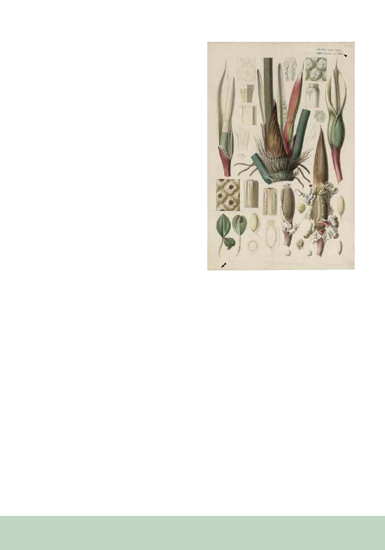
One Step Beyond: The Field
Drawings of Ferdinand Lucas Bauer
Use of illustrations as types is facilitated if they
were made using standardized coloration. An
outstanding example of this is the work of
Ferdinand Lucas Bauer (FLB), whose pencil field-
sketches, now almost all at NHMW, were the bases
for colored illustrations (mostly now at BM), some
of which are types. The drawings are important,
because they include information omitted from
the final, colored illustrations, such as collection
localities, dates, and additional detailed sketches of
descriptive morphological characters (Mabberley,
2021). The sketches also bear FLB’s notations using
numerical and other codes, which indicate the hue,
brightness, opacity, and texture of particular parts
of the living organism (Figure 2). FLB did this to
document rapidly the coloration of a specimen in
the field with a view to his “reviving” this later in
watercolor.
Figure 1. Note that this figure can only be accessed elec-
tronically via https://doi.org/10.5281/zenodo.7874857.
The lectotype for Philodendron imperiale Schott, a
synonym of Philodendron ornatum Schott (Araceae),
‘Schott Aroideae’ No. 3620 (see arrowhead). Most of
Schott’s specimens were destroyed in a fire at the end of
WWII, and taxonomists use the illustrations of original
material commissioned by Schott (this one done by W.
Liepoldt, double arrowhead) for typification.
FLB’s drawings recorded for the first time many
Australian species then new to Western science, as
he accompanied Matthew Flinders’ voyage (1801–
1803), the first documented circumnavigation
of the continent. For example, Figure 2 shows
an as-yet-undescribed species of Thomasia, a
genus restricted to Australia (Malvaceae). This is
likely T. sp. Vasse (Wilkins and Shepherd, 2019;
but see Shepherd and Wilkins, in preparation).
Exceptionally, the notation on the drawing
indicates that FLB drew it at Kew Gardens,
where the plant was raised from seed, most likely
collected during the expedition.
172
Figure 2. Drawing by Ferdinand Lucas Bauer of an undescribed species of Thomasia currently listed as T. sp. Vasse
(Malvaceae). Note that the numbers on the drawing correspond to numerical codes Bauer used to indicate hue, bright-
ness, opacity, and texture of a particular structure. The arrows point to additional symbols, such as sigils.

173
Color Charts for Standardization
in Historical Illustration as
Used by Bauer
FLB used the “painting by numbers” technique
throughout his career (Mabberley, 2017). His
color-code evolved over time and could have
been derived from principles in, or partial use
of, color systems developed by others. These
include Brenner (1680); Estner (1794), which is
specifically referenced on some of FLB’s Pacific
drawings (Mabberley, 2017, 2019); Schäffer (1769);
Schiffermüller and Denis (1771); Struve (1797);
Werner (1774); Widenmann (1794); Willdenow
(1799); and, perhaps, that associated with Haenke
(see Lack and Ibáñez Montoya, 2004, but also
Mabberley and San Pío Aldarén, 2012). FLB may
also have utilized color tables used by various
tradespeople (ceramics painters, printers, tanners)
and chemists (Pörner, 1773; Gülich 1779, 1780,
1781, 1786). Over time, he used an increasing
range of numbers, and the later work, in Australia,
included values close to 1000 (Pignatti-Wikus et
al., 2000) for botanical subjects, and beyond in the
later zoological drawings made on Norfolk Island.
However, FLB’s elaborate color chart is lost (Riedl-
Dorn and Riedl, 2019), if it ever existed (Mabberley
and San Pío Aldarén 2012; Mabberley, 2017, 2019,
2022; Jelley, 2022). Jelley (2022) has a practical
explanation for the numerical code based on an
art practitioner’s process, showing that the layout
of FLB’s paintbox may have functioned as his aide-
memoire and that the numbers could have been a
two-part code. The first one or two digits specified
which pigment to use, and the last digit(s) were
directions on how to achieve the correct blend,
brightness, opacity, etc. In other words, if this
hypothesis is proved, there probably never was a
physical color-chart of FLB's.
In addition to numbers, FLB used many additional
ciphers, especially in his later zoological drawings.
These include planetary symbols to convey
information about color, in that they were then
commonly understood to correspond to metals
such as gold for the sun ( ), silver for the moon
( ), and so on. However, FLB sometimes also wrote
the actual words “gold” or “silver” on drawings
together with the symbols. We assume therefore,
that the symbols had additional meaning and
may also refer to particular color blends, pattern,
texture, brightness, and/or opacity for that color
(e.g., yellow for ). Bauer also used upper- and
lower-case letters, maybe corresponding to those
in the tables of Schiffermüller and Denis (1771),
but also see Pignatti-Wikus et al. (2000), sigils
(symbols used in alchemy and magic; see Figure
2), and the Greek alphabet. Roman numerals
usually indicate the number of any particular
structure (e.g., stamens in a flower, spines of a fish
fin). It is currently mostly unclear, though, what
these additional symbols exactly mean. Jelley
(pers. comm.) is conducting further research into
these more complex layers of information in FLB’s
Australian work.
The importance of Ferdinand Bauer’s drawings at
the NHMW to taxonomic biology, as “iconotypes”
and otherwise, is only now beginning to become
clear. A thorough investigation of the collections,
necessarily involving international collaboration,
is much needed (Mabberley, 2021). In addition
to their immeasurable scientific and aesthetic
value, these drawings document the Australian
flora largely before European settlement. It would
therefore be important to make them available
to a broader audience. This will require funding
for a collaboration between Australian botanists,
NHMW staff (archive, botany, and library), and to
hire project-based digitization staff.
ACKNOWLEDGMENTS
We thank the reviewers for helping to improve
the manuscript, as well as Trevor L. Blake, Kelly
Shepherd (PERTH), and Carol Wilkins (PERTH)
for refining our Thomasia identification.

174
DATA AVAILABILITY
STATEMENT
High-resolution scans of the illustrations shown
and other “iconotypes” are available upon request
for research purposes via the Archive for the
History of Science at the Natural History Museum
Vienna (archiv@nhm-wien.ac.at).
CONFLICT OF INTEREST
The authors declare no potential conflict of
interest.
FUNDING
This project was not supported by external
funding.
AUTHORSHIP
S.F.: review and editing (equal); M.K.: review and
editing (equal); D.J.M.: conceptualization (equal),
writing – review and editing (supporting);
H.R.: review and editing (equal); M.-D.R.:
conceptualization (equal), writing – review and
editing (supporting), figure preparation (lead);
T.M.S.: conceptualization (equal), writing –
original draft (lead), writing – review and editing
(lead), figure preparation (supporting).
REFERENCES
Brenner, E. 1680. Nomenclatura et species colorum miniatae
picturae. Stockholm, Sweden. Website: https://weburn.kb.se/
metadata/146/EOD_2514146.htm
Coelho, M. A. N. 2000. Philodendron Schott (Araceae): mor-
fologia e taxonomia das espécies da Reserva Ecológica de
Macaé de Cima - Nova Friburgo, Rio de Janeiro, Brasil. Ro-
driguésia 51: 21–68.
Estner,
F. J. A. 1794. Versuch einer Mineralogie für
Anfänger und Liebhaber nach des Herrn Bergcommissions-
raths Werner’s Methode, I. Bd. J. G. Oehler, Wien, Austria.
Website: https://www.digitale-sammlungen.de/de/view/
bsb10283387?page=9
Gülich, J. F. 1779. Die rechte und wahrhafte Färbekunst. C. F.
Schneider, Leipzig, Germany. DOI:
https://doi.org/10.11588/
diglit.27273#0005
Gülich, J. F. 1780. Vollständiges Färbe- und Blaichbuch
zu mehrerm Unterricht, Nutzen und Gebrauch für Fabri-
kanten und Färber. A. L. Stettin, Ulm, Germany. Website:
https://books.google.at/books?id=zBVaxkKMGPoC&pri
ntsec=frontcover&hl=de&source=gbs_ViewAPI&redir_
esc=y#v=onepage&q&f=false
Gülich, J. F. 1781. Vollständiges Färbe- und Blaichbuch wel-
ches drey der wichtigsten Hauptstücke für Fabrikanten ent-
hält. Stettinische Buchhandlung, Germany. Website: https://
www.digitale-sammlungen.de/de/view/bsb10304876?pa-
ge=7
Gülich, J. F. 1786. Vollständige bewährte praktische Anwei-
sung zur Färberey auf Schaafwolle, Camellhaar und Seyde.
Stettinische Buchhandlung, Ulm, Germany. Website: https://
www.digitale-sammlungen.de/de/view/bsb10304882?pa-
ge=1
Jacquin, N. J. von. 1776. Florae Austriacae sive plantarum
selectarum in Austriae archiducatu sponte crescentium, Vol.
IV. J. M. Gerold, Wien, Austria. Website:
https://www.biodi-
versitylibrary.org/item/9678#page/7/mode/1up
Jarvis, C. 2007. The art and science of typification. In Order
out of Chaos. Linnaean plant names and their types, 13–61.
The Linnean Society in association with the Natural History
Museum London, London, United Kingdom.
Jarvis, C. 2008. Linnaeus’ use of illustrations in his naming
of plants. In M. J. Morris and L. Berwick [eds.], The Linnae-
an legacy: three centuries after his birth. The Linnean Special
Issue 8: 75–84.
Jelley, J. 2022. A Puzzle in a Paintbox: A painter’s solution
to Ferdinand Bauer’s colour code for the Flora and Fauna
Graeca 1786–1794. Art and Perception. DOI: https://doi.
org/10.1163/22134913-bja10042
Lack, W. and M. A. Ibáñez Montoya. 2004. Recording co-
lour in late eighteenth century botanical drawings: Sydney
Parkinson, Ferdinand Bauer and Thaddäus Haenke. Curtis’s
Botanical Magazine 14: 87–100.
Mabberley, D. J. 2017. Painting by numbers: the life and
work of Ferdinand Bauer. NewSouth, Sydney, Australia.
Mabberley, D. J. 2019. Botanical Revelation – European
encounters with Australian plants before Darwin The Peter
Crossing Collection. NewSouth, Sydney, Australia.
Mabberley, D. J. 2021. The “London” Australian natural his-
tory drawings of Ferdinand Bauer (1760–1826) revisited.
Flora Mediterranea 31: 67–94.
Mabberley, D. J. 2022. The Peter Crossing Collection: an il-
lustrated catalogue. Peter Crossing Collection, Sydney, Aus-
tralia.
Mabberley, D. J. and M. P. de San Pío Aldarén. 2012.
Haenke’s Malaspina colour-chart: an enigma. Real Jardín
Botaníco, CSIC, Madrid, Spain

175
Pignatti-Wikus, E., C. Riedl-Dorn, and D. J. Mabberley.
2000. Ferdinand Bauer’s field drawings of endemic Western
Australian plants made at King George Sound and Lucky
Bay, December 1801 - January 1802. I: Families Brassica-
ceae, Goodeniaceae p.p., Lentibulariaceae, Campanulaceae
p.p., Orchidaceae, Pittosporaceae p.p., Rutaceae p.p., Styli-
diaceae, Xyridaceae. Rendiconti Lincei, Scienze Fisihce e
Naturali 9: 69–109.
Pörner, C. W. 1773. Chymishce Versuche und Bemerkungen
zum Nutzen der Färberkunst. Vol. 3, M. G. Weidmanns Er-
ben und Reich, Leipzig, Germany. Website: https://books.
google.at/books?id=e-cUAAAAQAAJ&pg=PP5&hl=de&s
ource=gbs_selected_pages&cad=2#v=onepage&q&f=false
Riedl, H. 1981. Families destroyed in World War II at the
Vienna Herbarium (W). Taxon 30: 727–728.
Riedl-Dorn, C. and M. Riedl. 2019. Ferdinand Bauer or Jo-
hann and Joseph Knapp? A rectification. The Gardens’ Bulle-
tin, Singapore 71: 123–142.
Schäffer, J. C. 1769. Entwurf einer allgemeinen Farben-
verein oder Versuch und Muster einer gemeinnützlichen
Bestimmung und Benennung der Farben. E. A. Weiß, Re-
gensburg, Germany. Website: https://nbn-resolving.org/
urn:nbn:de:bvb:355-ubr12318-3
S
chiffermüller, I., and M. Denis. 1771. Versuch eines Farben-
systems. A. Bernardi, Wien, Austria. Website: https://www.
digitale-sammlungen.de/de/view/bsb10058712?page=1
Struve, H. 1797. Méthode analytique des fossiles, fondée
sur leurs caractères extérieurs. Lacombe & Co., Lausanne,
Switzerland. Website: https://www.digitale-sammlungen.de/
de/view/bsb10284666?page=1
Werner, A. G. 1774. Von den äußerlichen Kennzeichen der
Fossilien. S. L. Crusius, Leipzig, Germany. DOI: https://
digi.ub.uni-heidelberg.de/diglit/werner1774
Widenmann, J. F. W. 1794. Handbuch des oryktognos-
tischen Theils der Mineralogie. S. L. Crusius, Leipzig,
Germany. Website: https://www.digitale-sammlungen.de/de/
view/bsb10284860?page=1
Willdenow, C. L. 1799. Grundriß der Kräuterkunde
zu Vorlesungen entworfen. Von Ghelensche Schriften,
Wien, Austria. DOI: https://www.biodiversitylibrary.org/
item/220233#page/5/mode/1up
Wilkins, C., and K. Shepherd. 2019. A key to the species
of Thomasia (Malvaceae: Byttnerioideae). Nuytsia 30:
195–202.

176
From the P SB Special I ssue on Art in the Botanical Sciences
As archives of historical plant specimens,
herbaria provide snapshots of environmental
landscapes across time that are continuously
accessed, recontextualized, and reinterpreted
through modern techniques. Moreover, specimen
imaging in herbaria has been key to recent global
digitization efforts that confer multiple benefits,
including facilitating taxonomic revisions and
enabling better access to biodiversity data (Soltis,
2017). Specimens also become discoverable to
non-traditional researchers, such as artists and the
interested public.
From 2019 to 2022, the University of Texas at El
Paso (UTEP) herbarium imaged and georeferenced
all its specimens (~50,000 records) from the
southwest US and Mexico, funded by two grants
(IMLS IGSM-245733 and NSF DBI1902078).
To celebrate these projects, largely conducted
by biology undergraduates, we developed the
exhibit “Where We Will Grow: Elsie Slater, Plants,
and Art” at UTEP’s Centennial Museum and
Chihuahuan Desert Gardens (Fall 2021–Spring
2022) to showcase the history, art, and science of
the herbarium. We chose to highlight Elsie Slater
Celebrating the Launch of the UTEP Virtual Herbarium by
Highlighting Contemporary and
Historical Art and Science of the El Paso Region
Mingna V. Zhuang
1,3
Nabil Gonzalez
2
Michael L. Moody
1
1
UTEP Biodiversity Collections, Biological Sciences De-
partment, University of Texas at El Paso, 500 W Univer-
sity Ave. El Paso, TX 79968
2
Art Department, University of Texas at El Paso, 500 W
University Ave. El Paso, TX 79968
3
Author for correspondence: mzhuang@utep.edu
(1871–1952), a self-taught botanist, because her
collections represented the earliest specimens
from El Paso held by our herbarium and because
her unique dual interests in both the arts and
the sciences. Slater was a writer and artist whose
works are held at UTEP (Centennial Museum
and C. L. Sonnichsen Special Collections), which
span the scientific (exceptional botanical art) to
the impressionistic. Drawing from Elsie’s diverse
interests, the UTEP Herbarium partnered with
the Art Department’s Drawing I Class taught by
Nabil Gonzalez to use specimens from Elsie’s
collections as inspiration for student art. Because
of COVID-19 restrictions, art students visited the
new digital herbarium (Project Page: https://arctos.
database.museum/project/10003615) rather than
the physical herbarium. Collections Manager
Vicky Zhuang met with the students via Zoom
during a class period to introduce the project.
She described the herbarium, provided examples
of how researchers used herbarium specimens,
and guided the students through how to use the
database to find images of specimens. Students
were then instructed by Nabil to reinterpret Elsie’s
point of view towards the botanical world and
the romance of life. Each student researched their
chosen specimens and subsequently created a
composition representing a page out of a journal.
The students were asked to restrict their search to
the El Paso area and to focus on Elsie’s collections
when possible. However, they were intentionally
not exposed to Elsie’s artworks, so they would be
inspired to develop their ideas independently and
with few limitations. With regards to the exhibit,
students were only told that their pieces would be
paired with Elsie’s works and specimens in a botany
177
and art-focused exhibit and were encouraged to
explore a variety of mixed media and concepts.
This project granted students the experience of Elsie's
creative world that blended botany and art. For
example, Elsie’s specimen sheets often had poetic
notes. When describing a Baileya multiradiata
(UTEP 6602), she noted, “…one of the brightest
gold flowers, very very beautiful for our city.” Digital
collections allowed students to thoroughly examine
these specimens by focusing on shape, form, color,
and texture. Artists take inspiration from nature
and the world around them (Flannery, 2013).
Everything they see, touch, and feel is an important
part of how artists understand and research the
creative process. Thus, the collaboration gave
art students a new perspective on the artistry
of scientific specimens through Elsie’s unique
collection.
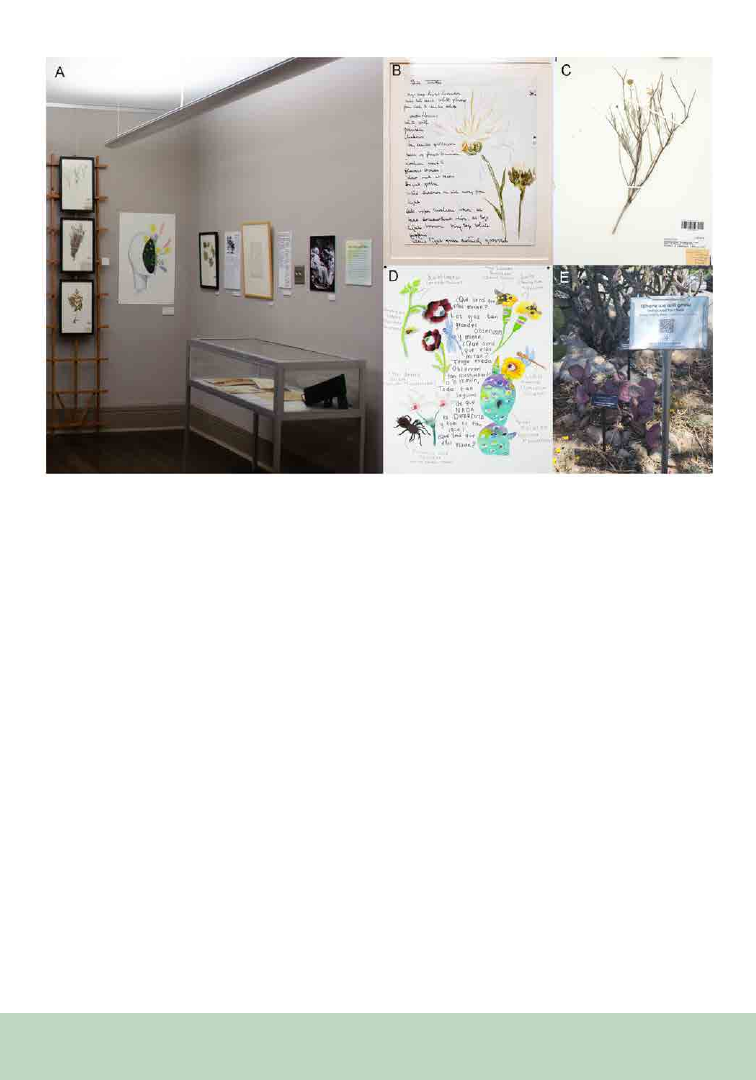
Because students were not restricted in style, the
students produced art pieces that represented
a diverse range of interpretations, realistic to
abstract, sometimes of the same specimens
(Figure 1). However, additional check-ins to
ensure the accuracy of scientific names and
species associations would have been beneficial.
Despite working independently, some students
converged on similar motifs (i.e., eyes, bones,
life, and death). Additionally, like some of Elsie’s
botanical art, some of the pieces included text (i.e.,
Figure 1B and D), as well as true-to-life reflections
of the specimens. Several art students found relief
from the pandemic lockdown and discovered new
inspiration in their local plant diversity. Finally,
biology students curating the exhibit connected to
the specimens they had been working on in a new
and broader context. Overall, the exhibit revealed
parallels in modern perspectives of science and art
using historical specimens.
Figure 1. Botanical-inspired art and science exhibited during the “Where We Will Grow Exhibit.” (A) A portion of the exhibit
pairing student art pieces with a description of Elsie Slater and her works, specimens, and art; (B) an example of Elsie’s art pieces;
(C) a Porophyllum scoparium specimen (UTEP 6646) used as art inspiration in part D; (D) art piece by Julyet Carillo, using sev-
eral species (UTEP 6646, 6696, 6649) showcased in the exhibit; (E) tour stop at the purple prickly pear (Opuntia macrocentra) in
the gardens, connecting living specimens to Elsie’s specimen (UTEP 6668). Photos courtesy of the Centennial Museum (A and B).

178
The resulting exhibit, “Where We Will Grow: Elsie
Slater, Plants, and Art,” combined Elsie’s botanical
specimens, art, and writings with diverse pieces
of contemporary student art. Curators selected
specimens from Elsie’s collections that were used
by students, represented in Elsie’s art or both
(i.e., Figure 1C and D). To draw cohesion with
the exhibit and living plants, patrons toured
the museum’s Chihuahuan Desert Gardens via
a self-guided tour app that linked living and
herbarium specimens. To choose the stops, we
cross referenced a list of specimens used by the
art students and species represented by Elsie’s
collections to species available in the Centennial
Gardens (i.e., Figure 1D and E). At each stop,
patrons scanned a QR code and viewed a specimen
collected by Elsie, its locality information, and a
few facts about the species. As a result, patrons
could view the same species in multiple contexts,
from the past, present, and future, through art, as
well as historical and living specimens.
The digital exhibit can be accessed at https://www.
utep.edu/centennial-museum/museum/past-
exhibits/where-we-will-grow.html.
REFERENCES
Flannery, M. C. 2013. The herbarium as muse: plant
specimens as inspiration. Biology International 53: 23-34.
Soltis, P. S. 2017. Digitization of herbaria enables novel
research. American Journal of Botany 104: 1281-1284.

179
From the P SB Special I ssue on Art in the Botanical Sciences
Alice V. Pierce
1,2,5,
Ian C. Anderson
1,3
Neelima R. Sinha
4
J. Grey Monroe
1
1
Department of Plant Biology, University of California,
Davis, Davis, CA, USA, 95616
2
Department of Plant Sciences, University of California,
Davis, Davis, CA, USA, 95616
3
Integrative Genetics and Genomics Graduate Group, Uni-
versity of California, Davis, Davis, CA, USA, 95616
4
Plant Biology Graduate Group, University of California,
Davis, Davis, CA, USA, 95616
5
Author for correspondence: avpierce@ucdavis.edu
ABSTRACT
Especially in recent decades, plant scientists
have had to develop new skill sets, becoming
statisticians, bioinformaticians, evolutionary
ecologists, visual artists, as well as experts in many
fields of biology. In regard to visual arts, botanists
have a long history of collecting plant specimens
for herbariums across the globe to showcase plant
diversity and through illustrations, they raise
awareness of the vast ecological importance of
plants in their diverse habitats. With botanical art,
plant scientists increase the public appreciation of
plant diversity and provide access to diversity in
regions where some of these plants had never been
seen. Now, with huge online repositories of digital
plant pictures, DNA/RNA sequencing, ChIP-seq,
metabolomics, and proteomic/crystallography
data, plant scientists have increased the ways to
catalog plant diversity at the molecular level, and
further increased the access of these resources
Celebrating Plant Diversity through Art
to fellow scientists. In this series of illustrations,
and through a modern digital twist of botanical
art, we hope to celebrate the progress made by
plant scientists around the world to accelerate our
understanding of plant evolution and diversity
and highlight a promising avenue for scientific
illustration to play a role in depicting fundamental
plant biological concepts and molecular diversity.
KEYWORDS
botanists, plant diversity, plant scientists, science
art
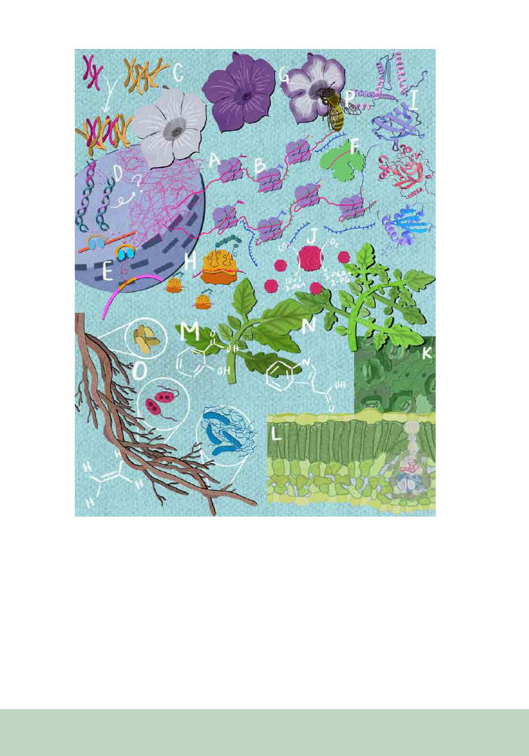
New technologies, such as DNA/RNA
sequencing, ChIP-seq, metabolite profiling, X-ray
crystallography, and others, have expanded our
methods for cataloging plant diversity by allowing
scientists to study plants at the molecular level.
In Figure 1, we celebrate the novel and impactful
efforts made by plant scientists to catalog
plant diversity across scales—from proteins to
Petunias. Understanding the diverse interactions
occurring at the molecular, cellular, organismal,
and environmental levels accelerates our
understanding of plant diversity and adaptation to
their environments.
Next to the chromatin, mRNA transcripts are being
subjected to RNA interference (F). At the top is an
homage to the petunia experiment where RNAi
was first discovered (G). Some of these transcripts,
however, can be transcribed by ribosomes and
become proteins (H). Many proteins interact at
the cellular level and work together to carry out
180
Figure 1. Several discoveries have revealed how plants interact and respond to their envi-
ronment. See the text of this article for further explanation.
a wide range of biological functions, including
transcription, translation, signaling, metabolic
processes (I), and carbon fixation (J).
On a larger scale, we see how cell types that have
differential gene expression patterns and vary in
protein populations can come together to form
tissues, such as in the stomata-epidermal cell
layer (K) and the leaf cross-section (L). These cells
interact with each other to communicate with
themselves and the environment to respond to
stimuli they might encounter.
Plants can respond to hormones such as auxin,
ethylene, and salicylic acid (M), which play a
crucial role in the diverse interactions occurring
within and between plants. Hormones regulate

181
plant growth, development, and response to
environmental and pathogen stress.
On the bottom, we see variation in tomato leaf
shape with age, known as heteroblasty (N). A
subset of phenotypic variation of developmental
traits allows plants to adapt and interact with their
environment differently, potentially providing an
advantage in various ecological niches depending
on the circumstances.
In addition to interacting with their abiotic
environment, plants interact with other
organisms. Understanding the interactions of
plants with other organisms such as microbes (O)
or pollinators (P) is critical for understanding the
role of plants in their ecosystem.
Continuing to catalog plant diversity at the
molecular, cellular, and environmental levels is
crucial to understanding and appreciating how
plants interact with their environment, furthering
our understanding of how plants have contributed
to the diversity of life, and accelerating efforts to
conserve plant species in a rapidly changing world.

182
From the P SB Special I ssue on Art in the Botanical Sciences
Brazilian Botanists Flirting with Arts:
Valuing the Multicultural Heritage
Lucas C. Marinho
1
Anderson dos S. Portugal
2
Vinícius dos S. Moraes
3
Marcelo G. Santos
2
Suzana Ursi
4
1
Universidade Federal do Maranhão, Departamento de
Biologia, Avenida dos Portugueses 1966, Bacanga, 65080-
805, São Luís, MA, Brasil.
2
Universidade do Estado do Rio de Janeiro, Faculdade de
Formação de Professores, Laboratório de Biodiversidade,
Rua Dr. Francisco Portela 1470, Patronato, 24435-005,
São Gonçalo, RJ, Brasil.
3
Instituto Oswaldo Cruz, Laboratório de Inovações em
Terapias, Ensino e Bioprodutos, Avenida Brasil, 4365,
Manguinhos, 21045-900, Rio de Janeiro, RJ, Brasil.
4
Universidade de São Paulo, Instituto de Biociências, De-
partamento de Botânica, Rua do Matão 277, Cidade Uni-
versitária, 05508-090, São Paulo, SP, Brasil.
5
Author for correspondence: lc.marinho@ufma.br
Plants have always been a source of inspiration
for many artists, and Botany often resorts to them
so that plants “live” eternally—as is expected—in
artistic works. And the plants, what can they “tell”
us about these humans who try to understand them
using Science and Art? Brazilian literature brings
rich examples in which plants are the protagonists
(see Clarice Lispector or Ana Martins Marques).
In the book O pensamento vegetal: a literatura e as
plantas (“Plant thought: literature and plants” in
free translation), Evando Nascimento, a Brazilian
writer, highlighted the relationship between
literary text and the floristic universe at the 19th
International Literary Festival of Paraty (Flip).
In this edition, the focus was on “Nhe’éry,” the
Atlantic Forest as named by the Guarani, one of
the many native peoples of Brazil.
Initiatives focusing on this powerful Art–Botany
relationship are still germinating in Brazil
(following “ArtScience Manifesto”; see Bernstein
et al., 2011), with some botanists and teachers
building upon the multisensory and poetic
experience created by plants and the world. It
is clear that, even though sparsely distributed
throughout Brazilian history, there have been
other non-scientific artistic initiatives between art
and plants. But here, we present some of the Art
and Botany (as a science) initiatives and, especially,
contextualize the complex cultural interplay that
shaped these two areas in Brazil.
COLONIAL HERITAGE
The arrival of Europeans represented a breaking
point in how art was seen and produced in Brazil.
However, there is a rich recorded Brazilian Pre-
Cabraline Art (in reference to the Portuguese
navigator Pedro Álvares Cabral, the “discoverer” of
Brazil), represented by cave painting, sculptures,
and ceramics. Amazonian ceramics are probably
the best-known artistic manifestations of
Brazilian Pre-Cabraline Art (Prous, 2007). Prior
to colonial contact, Indigenous art was sovereign;
subsequently, other cultures, especially from the
African continent, also set out their point of view.
The art of the remaining Indigenous peoples is still
quite expressive in Brazil (for more, see examples
of Carmézia Emiliano and Uýra Sodoma). The
presence of artists (e.g., Thomas Ender and Johann

183
Moritz Rugendas) in the field expeditions to the
interior of Brazil showed the European vision of
the Brazilian flora, and everyday aspects for the
original people took on grandiose dimensions for
those who were unfamiliar with it.
Botany assumed the status of Scientia Amabilis
at the end of colonial period in Brazil, when the
knowledge about plants was seen as an important
social status with reverberations in architecture, in
gardens, and in the great scientific expeditions to
recognize the flora and the associated biodiversity.
National Botany Day in Brazil celebrates the
birthday of a Bavarian naturalist, Carl Friedrich von
Martius. Although this symbolic date highlights
how the European view is still hegemonic in
Brazilian Botany, it is impossible to ignore the
tremendous value of Martius’ legacy. Together
with Johann Baptist von Spix, Martius traveled
immense distances through Brazil between 1817
and 1820 throughout a variety of domains and
recorded all the floristic diversity they encountered
in the magnificent Flora Brasiliensis compendia.
They returned to Europe with a huge collection of
preserved biological samples and live specimens.
Two young Indigenous people from different
ethnicities in Brazil, Miranha and Juri, were also
taken to Europe and died some time later. The
story of how those people were integrated into
Martius and Spix’s expedition is still controversial
and has more than one version (Costa, 2019).
At the end of his life, Martius rejected the brutal
behavior of including people as collectibles, which
was sadly common among colonizing naturalists.
Martius and Spix produced valuable ethnographic
descriptions, and the masks collected by them in
Amazonia are an important record of symbolic
practices by Indigenous nations, many of them
now extinct (Santos, 2014).
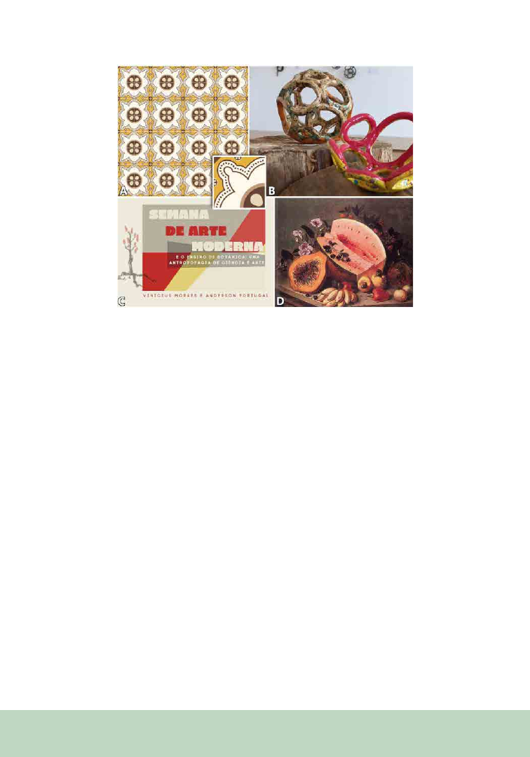
Gradually, not only was European culture brought
to Brazil, but also species in vivo or in graphic
representations, such as those that adorn the
Portuguese tiles, or azulejos, of northeastern Brazil
(Menezes et al., 2020). In this case, it was up to
Brazilians to appreciate the Europeans’ paintings
of non-native plants without the feeling of cultural
identity (belonging) about the artifact or what it
represented (Silva et al., 2021).
Brazilian flora began to be included slowly in
art as part of sacred works and sculptures (e.g.,
Machado et al., 2018). At that time, art in general
was closely linked to Catholic productions, and
much of what was produced had underlying
tendencies. In this sense, a devaluation of the
arts produced by cultural groups that existed
in Brazil (such as that by Indigenous people) or
that were brought to Brazil (such as by Africans)
was inevitable, since until then these local groups
did not share the Christian faith. Although the
quality and historical value contained in the
Portuguese azulejos and sacred sculptures are
invaluable, the presence of plants is linked to the
technical character, as part of the work, and not as
something to be felt (Figure 1).
Contrary to the visual arts, music and dance had
already been impacted by the presence of different
cultures on Brazilian land. Orality carried out
information about plants from north to south of
the country. For example, in Samba or Capoeira
(a dance/fight created by Brazilians of African
descent) songs, the use of native plants in the
production of percussive instruments and rituals
is common (Hartmann et al., 2023). The fact is
that, although the Brazilian population of the
colonial period was composed of many people—
it is important to highlight that Indigenous and
Africans were composed of countless different
ethnic groups, and each one had its own way
of seeing art and plants—the contribution for
“botany and art” was unbalanced.
Decolonization Movements
In 1922, a great artistic exhibition called “Semana
de Arte Moderna (SAM)” took place in São
Paulo. Organized by artists and intellectuals,
the movement suggested breaking up with
artistic European traditions of the time. SAM is
184
considered a symbolic reference of the beginning
of Brazilian Modernism period. One of the works
that had a great impact on its configuration was
the Manifesto Antropofágico, published in 1928 by
Oswald de Andrade. The document proposes a
process of swallowing European ideas, techniques,
and cultural thoughts to transform them, without
being submissive, but critical, in the promotion
of Brazilian art in essence (Ajzenberg, 2012). The
representations of Brazilian flora in modernist
productions are vast and present the great
Brazilian biodiversity. Ribeiro (2020) reports the
mention of more than 200 plant species in these
works of art. In her book, she discusses 19 of these
plants, such as banana, cactus, corn, coffee, and
Brazil wood. Moraes and Portugal (2021) reflect
on the use of some of these species in teaching
processes from a decolonial perspective.
Although it has great importance in the country’s
historical and cultural scene, Brazilian Modernism
is not marked as a genuine Brazilian movement.
In the 1920s, the coffee-growing elite (known to
be former enslaved peoples’ owners) financed
SAM. The art movement was built by and for the
Brazilian elite, without considering the knowledge
and cultures of other populations living in a
country as large and diverse as Brazil.
Less prominent than SAM, the Movimento
Armorial (MA) was a relevant artistic initiative for
the appreciation of popular knowledge. MA started
in 1970 and was led by playwright and writer
Ariano Suassuna (1927–2014). The movement
aimed to produce an authentic Brazilian art that
is erudite but rooted in Brazilian popular culture.
The movement guided the convergence of artistic
expressions to produce a national art, having
Figure 1. (A) Integration between Botanists and Designers to create new prints for “azulejos” in Maranhão using
native species (an “azulejo” of babaçu [Attlaea speciosa Mart. ex Spreng.]; see Silva et al., 2021). (B) Artwork detail
of the exhibition “Weavings of life - Art and Botany,” at Science and Technology Park of the University of São Paulo
(described by Ursi et al., 2023). (C) Short course offered by Moraes and Portugal on the representation of botany in
works of art of Brazilian modernism and its possibilities of use in teaching. In these materials, there is the possibility
of educational work on: monocultures, different land uses and environmental degradation, capitalist mode of produc-
tion, exclusion of socially minority groups, and the production of paints from coffee fruits and these products. (D)
Natureza-morta (1868; Agostinho José da Mota). Painting used in a guidebook of daily plants (Marcelo G. Santos,
2023 in press) to discuss native and exotic fruit. Photo in (B) by T. Cesquim.

185
the northeastern region of Brazil as a source of
inspiration. In these productions, the Caatinga
domain was portrayed amidst the experiences of
its people. Such initiative arises from the need
to value local productions and northeastern
scenarios, in which the great biological and social
diversity of the Caatinga is highly valued.
The counterpoint to this thought is the production
of Literatura de Cordel (LC), which uses popular
language with rhymes and verses that present
metric perfection. Despite its colonial heritage, LC
is an artistic expression intimately linked to the
Brazilian northeast. LC portrays the knowledge
and experiences of the people of the Northeast.
LC, rich in images produced through woodcut
and use of a clothesline for its public exhibition,
highlights Brazilianness. Because of this, it is used
as a didactic tool by several educators, including
in the teaching of Botany. For more on LC and
Botany, see Oliveira and Cavalcante (2020) and
Santos et al. (2022).
Embrionary Botany
Art Initiatives
From the Martius expedition until now, some
Brazilian botanists seek to reconnect Scientia
Amabilis with art, but from a new perspective,
valuing the diversity of environments, cultures,
ethnicities, and regional characteristics (Figure 1).
These efforts of botanists in Brazil to merge plants
and art have been successful. However, they are
still stuck in a utilitarian metric—always trying
to answer “what is this for?”—that underuses the
driving force of art. We have a long way to go to a
ScienceArt expression that promotes diversity, but
starting this conversation is the first step to walk
this path.
ACKNOWLEDGMENTS
We thank Dr. Charles C. Davis (Harvard
University) for critical reading of an earlier
version of the manuscript. We are also indebted to
the reviewers who carefully reviewed our article.
REFERENCES
Ajzenberg, E. 2012. A semana de arte moderna de 1922. Re-
vista de Cultura e Extensão USP 7: 25–29.
Bernstein, R., T. Siler, A. Brown, and K. Snelson. 2011. Art-
Science: integrative collaboration to create a sustainable fu-
ture. Leonardo 44: 192.
Costa, M. F. 2019. Os “meninos índios” que Spix e Martius
levaram a Munique. Artelogie 14: 1–17.
Hartmann, J. S., E. M. Schneider, and L. Biral. 2023. Princi-
pais espécies vegetais usadas na capoeira. Botânica Pública 4:
12–18.
Machado, P. F. S., J. N. C. Marchiori, and D. Sanches. 2018.
Anatomia do lenho de esculturas do Museu Vicente Pallotti
(Santa Maria, RS, Brasil). Balduinia 63: 1-19.
Menezes, L. R., A. B. Ewerton, A. L. Garcia, S. S. Dominici, F.
R. Fernandes, L. F. A. Campos, and L. C. Marinho. 2020. The
Flora of Azulejos of Maranhão, Brazil. Ethnobiology Letters
12: 94–102.
Moraes, V. S., and A. S. Portugal. 2021. Obras Modernistas e
a Botânica: A construção de uma brasilidade e suas possibili-
dades de ensino decolonial. Vitruvian Cogitationes 2: 74–87.
Oliveira, R. L. C. and W. A. Cavalcante. 2020. Cordel para
o Ensino de Botânica: Morfologia, 2. ed. Boa Vista: UERR
Edições.
Prous, A. 2007. Arte pré-histórica do Brasil. Belo Horizonte:
C/Arte.
Ribeiro, A. C. C. 2020. Pequeno guia da botânica modernista.
São Paulo: Editora da Autora.
Santos, M. S. 2013–2014. Naturalists in Nineteenth-Century
Brazil. Archiv Weltmuseum Wien 63–64: 38–59..
Santos, A. C. R., E. N. R. Moreira, F. M. Aragão, G. S. Silva,
J. S. Sousa, and M. M. S. Castro. 2022. Botânica em poesia:
fotossíntese e respiração numa perspectiva cultural. Botânica
Pública 3: 7–12.
Silva, K. L. B. M., L. C. Marinho, F. R. Fernandes, and L. F. A.
Campos. 2021. O patrimônio azulejar de São Luís e a flora
maranhense: uma proposta conceitual para a criação de es-
tampas com uma identidade regional. Anais do 10º CIDI e
10º CONGIC.
Simões, A. 2022. Reapropriar para reparar: o centenário da
Semana de 22 sob a ótica decolonial. Revista USP 135: 215–
228.
Ursi, S., D. Amaral, and T. Cesquim. 2023. Exposição “Tra-
mas da Vida - Arte e Botânica”: encantamento e conhecimen-
to no Parque CienTec da USP. In: B. R. S. Cerqueira, L. C. A.
B. Souza, A. Pugliese, D. M. S. Medeiros, V. H. O. Henrique
(Orgs). E a vida continua: vazios e esperanças no Ensino de
Ciências e de Biologia: Coletânea de trabalhos do V EREBIO-
R1, São Paulo: Pimenta Cultural, pp. 263-272.
186
From the P SB Special I ssue on Art in the Botanical Sciences
Nezka Pfeifer
Museum Curator,
Stephen and Peter Sachs Museum,
Missouri Botanical Garden
Botanical gardens create a unique opportunity
to intersect science, culture, and art to offer
interdisciplinary experiences for the public. The
Missouri Botanical Garden’s historic (ca. 1859)
Botanical Museum was restored, renovated, and
renamed the Stephen and Peter Sachs Museum
and reopened to the public in late April 2018
after more than a century of alternate use. As
the Museum Curator and sole staff member
responsible for the Sachs Museum, I curate
exhibitions using botanical science as the focus
of the exhibition narrative, interweaving history,
material culture, and inclusive perspectives,
together with commissions of contemporary site-
specific work. As a part of the Science & Research
Division at the Garden, the Sachs Museum
collaborates with departments managing other
plant-based collections, including the Herbarium
(dried plant specimens) and the William Brown
Integrating Botany, History, Culture, and
Contemporary Art in a Botanical Garden Museum
Center’s biocultural (ethnobotany) collection, to
feature the important work Garden staff is doing
in botanical science.
Visitors to the Missouri Botanical Garden come
from a spectrum of backgrounds—some are well-
versed in botany and horticulture, but many are
not. The challenge is to engage these publics on
the myriad ways plants are relevant to our lives, yet
also include the botanical science that the Garden
experts work on daily in St. Louis and around the
world. I focus on subjects that embrace the ubiquity
of plants, so that visitors enjoy the rediscovery of
plants on a subject previously overlooked. The
foundation of the exhibition subject is on botany
(and horticulture and entomology when relevant);
this topic might feature in Garden research or have
a universal impact and appeal. Where possible,
I include little-known connections from other
disciplines and collaborate with other science and
community organizations, museums, and lenders
to highlight information that might impact
visitors on a personal level, such as regional
history or material culture. Finally, I commission
contemporary artists to create artworks that
interpret the botanical subject in unique and
meaningful ways to expand understanding about
the subject. The two most recent exhibitions at
the Sachs Museum embodied the key goals I
am trying to achieve with this multidisciplinary
framework. I also create live in-person programs
as well as digital content for exhibitions for both
education and promotion; this includes musical
performances, virtual tours, multiple blog posts,
myriad social media posts on X (Twitter) and
Instagram, and talk series highlighting the art and
science connections.
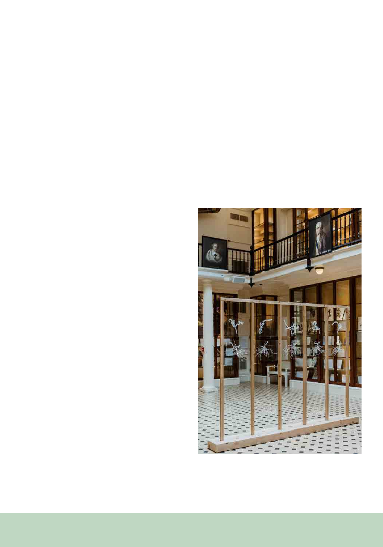
187
To celebrate Missouri’s bicentennial of statehood
in 2021, I planned an exhibition to focus on a
Missouri viticultural innovation with deep roots
in the botanical history of the state and long-
lasting impacts on viniculture around the world.
The exhibition intertwined this history with
contemporary scientific research investigating this
innovation through several site-specific artworks
focused on the grapevines using contemporary
media of film and machine learning. Grafting
the Grape: American Grapevine Rootstock in
Missouri and the World explored the millennia-
long human cultivation of wine from the grapes
of Vitis vinifera as a drink for social, religious,
and economic power. The native American
grape species that were used historically by the
Indigenous peoples of North America were also
used in the viticulture of early colonial Missouri,
and continue in the state’s wine industry today.
In order to address a global insect infestation in
the 1860s, Missouri scientists and horticulturists
grafted these native rootstocks with the Vitis
vinifera grape scion, thereby enabling the survival
of the wine grape. Without the research and
innovation by 19th-century Missouri botanists,
entomologists, and viticulturists, drinking wine
from this species would not be possible today.
Artist Dornith Doherty—renowned for her
photographic work on seeds and global seed-
banking—created photographs and two short
films for her series Roundabout (Circuition)
inspired by the intersection of the historical
innovation and the contemporary scientific
research taking place in Dr. Allison Miller’s NSF-
funded Vitis Underground project (exploring
the impact of different rootstock species on the
grafted grape berry). Artist collaborators inspired
by the impact of climate change on grapevines,
Lei Han and Lorraine Walsh focused their work
for Grafting the Grape on environmental shifts as
seen through the seemingly disparate practices
of ancient horticultural grafting techniques and
contemporaneous machine learning (a subset
of artificial intelligence). Their art focused on
the native grapevine species Missouri Vitis
aestivalis (also known as Missouri’s Norton grape)
and the process of this mediation in order to bring
a fruitful awareness of the significant effect climatic
change has on life. They created drawings, digital
images, and three short films for the exhibition,
as well as a large sculpture titled The In-Between
(Figure 1). This stylized wooden trellis held two
horizontal rows of five plexiglass plates each, with
the upper row featuring engraved drawings of
the grapevines and the lower row engraved with
drawings of the rootstocks; the negative space in-
between the plexiglass plates is where the grafting
occurs, and the viewer’s imagination fills in the
blank. Visitor feedback consistently was one of
awe; for the beauty and character of the artworks
on display, but also for the global impact of this
integral innovation that is still relevant today.
Figure 1. The In-Between, abstract grafted grapevine
sculpture, by Lei Han and Lorraine Walsh, 6’6”L x 1.5”D
x 8’9”H, ca. 2021. (Photo credit: Virginia Harold)
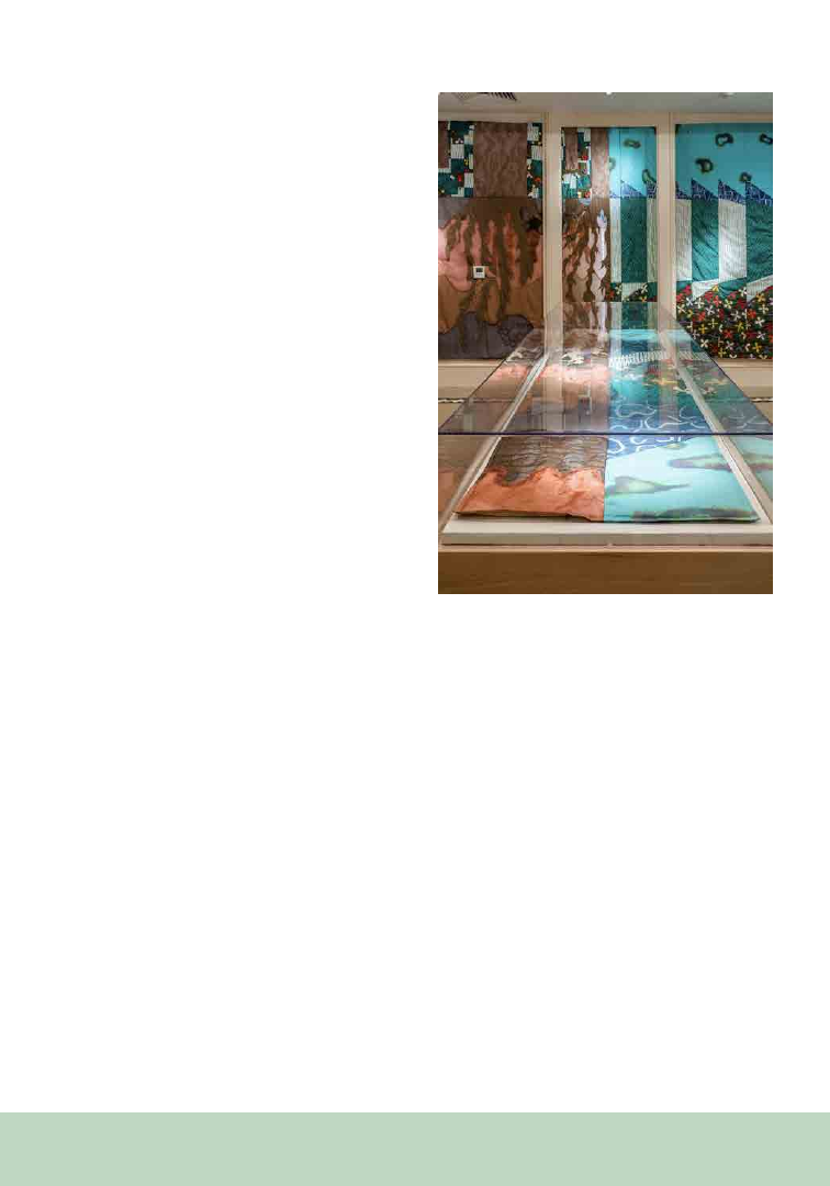
188
The 2022 exhibition Botanical Resonance:
Plants and Sounds in the Garden embraced how
important plants are in the human soundscape—
both in nature and culture—and how these species
are endangered globally due to overharvesting
and climate change. A particular focus included
how the Garden is working with scientists and
musical instrument makers around the world
to address some of these conservation issues.
In conversation, visitors and other curators
continually remarked on how they were previously
unaware of how dependent humans are on plants
to create sound and music. A series of live music
performances featuring plant-based instruments,
such as alphorns and west African idiophones,
gave opportunities to experience (hear, feel,
sense) the vibrations of sound making in person.
I commissioned three contemporary artists to
create artworks interpreting plants and sounds
in different ways. Annika Kappner created two
auditory guided soundwalks (accessed via QR
code) that provided a meditative exploration of
plants and sounds into the Garden. Brooke Erin
Goldstein designed a full gallery quilted room
installation visualizing the ways plant families
communicate with one another, in which the
world is bisected, above and below (Figure 2).
As pictured, one half of the gallery featured a
forest’s underground root system network, and
the other half illustrated a manicured yard of
grass that had just been cut, visualizing the grass
screaming (we usually experience this as the smell
of cut grass). Kevin Harris composed and built an
electronically synthesized immersive rainforest
sound installation as an offering to the botanical
world that nourishes us every day. A virtual talk
series with the artists gave detailed presentations
on the creative process for the artworks in the
exhibition.
Curating exhibitions to intersect plant science,
human culture, and art expands the possibilities of
interpretation for the public, opening up avenues of
engagement that make botanical content relevant
for many visitors who might be intimidated by
science-only content. While anecdotal and not
quantitative, feedback from my conversations
with the public and other curatorial colleagues has
made clear that approaching a subject of popular
interest—while including botanical science and
art—makes an impact on how people understand
(and enjoy learning about) how connected plants
are to our lives. These experiences remove barriers
to information and build connections that will
lead to deeper understanding of plants, as well as
the larger world around us.
Figure 2. Reverberations, an immersive quilted room
installation by Brooke Erin Goldstein, 26’4”L x 19’10”D
x 8’4”H, ca. 2022. (Photo credit: Virginia Harold)

189
From the P SB Special I ssue on Art in the Botanical Sciences
The raw form of scientific data is usually
unfamiliar to a general audience. One exception
to this are photographs. Lenses and cameras are
common tools in scientific, artistic, and popular
use, and the photograph represents a rare example
of a shared medium across these contexts. For
the scientist, a photograph is raw data (e.g., a
scanning electron micrograph of pollen grains
on a stigmatic surface or an in situ hybridization
assay of gene expression in a developing leaf). For
the artist, though, a photograph is an expressive
object produced by their creative work. In both
cases, the photograph is a visual record of a
lens pointed at a subject, regardless of whether
the primary intention is analytical or aesthetic.
This is a remarkable confluence of two different
approaches to learning about the world (science
Science, Art, and the Allure of Photographs
Benjamin Goulet-Scott
1,2,4
Jacob S. Suissa
1,3,4
1
Let’s Botanize, Inc. Arlington, MA, USA
2
Harvard Forest of Harvard University Petersham, MA,
USA
3
Department of Ecology and Evolutionary Biology, Uni-
versity of Tennessee at Knoxville, Knoxville, Tennessee,
USA
4
Pratt Institute Brooklyn, NY, USA
and art) that are often regarded as wholly distinct
fields. Here, we explore the photograph and the
cultural intersection between science and art,
asking what consequences and opportunities arise
from the shared practice of lens-based image-
making across these two disciplines.
Science and art are both concerned with producing
insightful observations of the world. Hand lenses,
microscopes, and cameras augment how we see—
focusing, enlarging, or warping a visual field. Our
view through the lens represents the world in a way
that reminds us of our natural perception but may
be impossible to achieve through unaided sight.
However, lenses do not generate an image from
nothing. Rather, they project only and exactly
what the photographer frames in front of them. We
say that photographs are taken—images plucked
directly from the fabric of reality. Recognizing
this, we imbue photographs with an objective
authority over the truth not granted to other visual
media. “More convincingly than any other kind of
picture, a photograph evokes the tangible presence
of reality. Its most fundamental use and broadest
acceptance has been as a substitute for the subject
itself—a simpler, more permanent, more clearly
visible version of the plain fact” (Szarkowski, 1966).
For artists, this makes photography unique from
other visual media, such as drawing and painting.
For scientists, this means that photographs can be
regarded as trustworthy primary data.
It is important to recognize that the status of
photographs as objects of truth has been abused
since the early days of the medium. We do, after
all, have photographic “evidence” of the Loch
Ness Monster, Bigfoot, and the Cottingley Fairies.
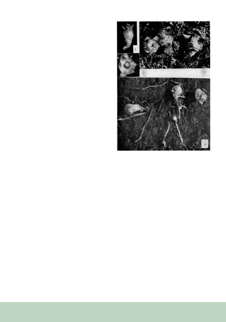
190
Digital technologies like Photoshop and generative
Artificial Intelligence further complicate the
photograph’s reputation for objective truth-telling.
Furthermore, our belief that cameras are objective
recording devices may lead us to downplay the
influence of a photographer’s biases and agendas,
which are expressed in a series of decisions about
composition, focus, and moment of capture. Yet,
our fundamental trust in photography endures,
even if it is now, more than ever, accompanied by
a healthy dose of skepticism.
Plants are particularly well-suited to photographic
observation because they do not move. In fact,
the first two books to be illustrated by early
photographic techniques both featured images of
plants. One of the pioneering women in botany,
Anna Atkins, created Photographs of British Algae:
Cyanotype Impressions (1843), which was entirely
devoted to plant subjects, and the 24 photographs
in William Henry Fox Talbot’s The Pencil of Nature
(1844) are primarily of architectural subjects with
the organic exceptions “Leaf of a Plant” and “A
Fruit Piece.” We can also look to plants for an
early example of the power of photography in
the scientific record. In 1912, botanist Dr. Norma
Etta Pfeiffer discovered a miniscule plant growing
within the city limits of Chicago, which was then
the second largest city in the United States. She
described this plant as a new species, Thismia
americana, recognizing that it belonged to a genus
of small mycoheterotrophic flowering plants that
had not been observed in North America before
(or since). Unfortunately, this plant has not been
found since 1916 and is presumed extinct due to
habitat loss. So how can we be sure that such an
unlikely plant even existed? Indeed, the possibility
of a botanical hoax devised by Dr. Pfeiffer has
been raised (see Wilhelm and Rericha, 2018).
However, in addition to Dr. Pfeiffer’s reputation
as an excellent botanist and plant morphologist,
the T. americana story is believed in part because
she took and published photographs of the plant
in 1914 (Figure 1; Pfeiffer, 1914). The suspicion
of a hoax would likely be much stronger had Dr.
Figure 1. Norma Etta Pfeiffer’s photographs of Thismia
americana published in 1914. These images confirm the
existence of this species, now thought to be extinct. (Im-
ages in the public domain.)
Pfeiffer chosen to publish drawings of the plant
instead of photographs.
Today, photography (still or moving) is a primary
mode of visual communication. The medium
is a democratic one—most people make and/
or consume photographs every day. Due to the
ubiquity of photographs and their relationship with
the truth, looking at a photograph is an intuitive
way for an audience to learn about the reality of
the world. This suggests that photography-based
platforms like most social media have enormous
educational potential (provided that the content
is unaltered and accompanied by credible
interpretation; Figure 2). The global popularity
of these platforms (exceeding 1 billion users)
attests to the ability of photography to grab our
attention and hold it. A photograph is memorable,

191
Figure 2. A soon-to-emerge leaf of Liriodendron tulip-
ifera is visible through large enclosing stipules. Visually
arresting photographs are powerful teaching tools when
accompanied by expert interpretation. This image was
originally posted to Instagram (@letsbotanize). (Photo-
graph by Jacob Suissa.)
expressive, and persuasive with minimal barriers
to engagement for broad audiences. Photography
is, therefore, one of the most powerful tools
available to educators, artists, and scholars aiming
to communicate credible information with large
audiences because it is both an aesthetically
engaging and data-rich medium.
The accessibility of photo-taking has also made
possible a tool like iNaturalist, which crowdsources
“research-grade” photographic data at a global
scale. The iNaturalist image database has been used
to train machine learning algorithms that attempt
to identify organisms through the live camera feed
of a smartphone. Interestingly, the capabilities of
such algorithms are determined by (and reveal)
trends and biases, including the way photos of
other species are composed, which phenological
stages tend to warrant photographing, which
populations use iNaturalist, and which habitats
are easily accessed by humans. These biases
occur regardless of which angles, features, and
habitats would be most taxonomically useful.
For instance, details of the bud and leaf scar are
very useful in distinguishing among species in the
hickory family (Juglandaceae), but rarely warrant
the photographic attention of non-botanists. In
theory, such biases and gaps should only diminish
over time as more images are added to the
training dataset. iNaturalist takes advantage of the
photograph’s dual identity as an object of beauty
and an object of data to engage broad audiences in
the practice of research.
As science communicators, we are particularly
excited about the unique potential of photography
to provoke curiosity and excitement about
the natural world. John Szarkowski describes
photographers as recognizing that “the world
itself is an artist of incomparable inventiveness”
and “the compelling clarity with which a
photograph recorded the trivial suggested that
the subject had never before been properly seen,
that it was in fact perhaps not trivial, but filled
with undiscovered meaning” (Szarkowski, 1966).
Everything in frame and in focus is recorded with
equal fidelity by a camera. Take a photo outside,
and the unfathomable complexity of the natural
world is all there at a level of intricate detail that a
human mind could never generate from scratch;
the only limit is the resolution of the photograph.
Plants especially benefit from this feature of
photography, since unlike people, cameras do
not experience plant awareness disparity (Parsley,
2020). Because plant life dominates terrestrial
biomass, most photographs of “nature scenes” will
contain plants (likely in abundance). Inevitably,
bird photography, snake photography, and insect
photography are also plant photography, even if
the photographer themself only considered the
plant life as patches of green to be balanced within
the composition. Fortunately for the botanist or

192
the plant-focused science communicator, those
plants lay in wait within the frame, ready to be
discovered as anything but trivial. The medium
of photography itself incites a sense of awe and
curiosity and a spark to explore the image in
search of beautiful moments. These are exactly
the emotions that science communicators hope to
inspire in their audience.
The photograph is both scientific and artistic;
composing and recording an image through a
lens is one instance in which the two practices
coalesce. Both endeavors value photography’s
capacity for creating information-dense and
captivating records of reality. Whether deployed
in science, art, or in braiding the two together,
photography wields enormous potential for
learning and communicating about the world.
Through photography, science communication
has the capacity to reach larger audiences than ever
on visual-driven social media platforms. At the
same time, deploying photography for scientific
research or education must be accompanied by
a deep sense of responsibility to not abuse our
intuitive trust of the medium.
REFERENCES
Atkins, A. 1843. Photographs of British Algae: Cyano-
type Impressions.
Parsley, K. M. 2020. Plant awareness disparity: A case
for renaming plant blindness. Plants, People, Planet 2:
598-601.
Pfeiffer, N. E. 1914. Morphology of Thismia ameri-
cana. Botanical Gazette 57: 122-135.
Szarkowski, J. 1966. The photographer’s eye. The Mu-
seum of Modern Art, New York.
Talbot, W. H. F. 1844. The Pencil of Nature. Longman,
Brown, Green and Longmans, London.
Wilhelm, G., and L. Rericha. 2018. Thismia Ameri-
cana: A Chicago Endemic or an Elaborate Hoax? The
Great Lakes Botanist 57: 150-157.

193
From the P SB Special I ssue on Art in the Botanical Sciences
ABSTRACT
PhyloPic is a crowd-funded database of organism
silhouette images that currently contains 8364
accepted submissions from 585 volunteers. Each
silhouette image is associated with a taxonomic
name via a network of phylogenetic nodes,
enabling dynamic searches based on evolutionary
relationships. The project has become an
invaluable artistic resource for scientific
publications, with more than 1900 references in
published articles. Despite its success, adoption
and contribution by plant scientists has been
relatively low. Only ~8% of current submissions
are for organisms within Archaeplastida (Plantae
sensu lato), representing just ~0.066% of named
terminal phylogenetic nodes (i.e., species or
Filling Out PhyloPic: Call for Adoption by
Plant Scientists
Mason C. McNair
1, 3
T. Michael Keesey
2
1
Clemson University, Pee Dee Research & Education
Center, 2200 Pocket Rd, Florence, SC 29506
2
PhyloPic, 22171/2 Addison Way, Los Angeles, CA
90041
3
Author for correspondence: mcnair5@clemson.edu
subspecies). To further improve the utility of
PhyloPic and promote increased use across the
plant sciences, we showcase fast and efficient
methods for creating suitable silhouettes from
multiple image sources including photographs,
illustrations, and herbarium specimens. These
methods will help increase adoption and quality
of submitted images and complements the recent
release of PhyloPic 2.0, which features faster
performance, an improved user interface, and
automatic conversion of submissions to infinitely
scalable vector files.
KEYWORDS
creative commons, figure creation, open access,
PhyloPic, scientific communication, silhouette
images
Creating impactful figures to effectively
communicate science is challenging. Many
scientists aspire to create educational and inspiring
figures but struggle due to a lack of artistic skills
or limited access to high-quality graphics. These
barriers can be eased through expansion of the
PhyloPic platform.
PhyloPic is an open database of free silhouette
images of organisms. Each silhouette is associated
with taxonomic names via a curated phylogeny
drawn from a variety of sources, primarily The
Open Tree of Life (OpenTreeofLife et al., 2019)
and The Paleobiology Database (McClennen et
al., 2017). This enables dynamic searches based
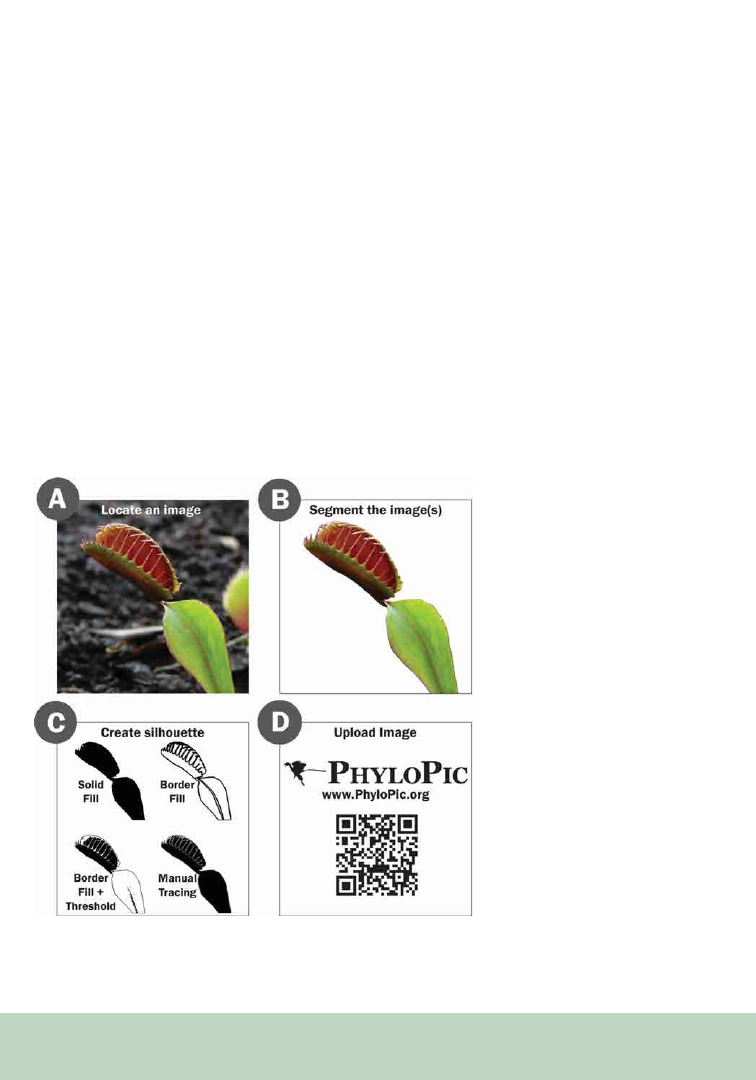
194
on phylogenetic relationships, either manually
through the website (https://phylopic.org) or
programmatically, through a public Application
Programming Interface (API; https://www.
phylopic.org/articles/api-recipes) or the R
package (Gearty et al., 2023). Despite the PhyloPic
platform being written in English, the binomial
nomenclature used for searching the website
is universal and enables use of the platform
regardless of a user’s language. Website users can
also create collections of silhouettes that can be
used to create permanent links for attribution. The
“Collections Drawer” tool enables users to create
named sets of silhouettes for later use. Collections
are private and stored in the user’s browser cache,
but they can be turned into permanent shareable
links.
Currently, PhyloPic features 8364 accepted
submissions from 585 volunteers. The project
has become an invaluable artistic resource for
scientific publications, with more than 1900
references in published articles. However, plant
taxa are noticeably underrepresented on PhyloPic,
with only 669 silhouettes, making up just ~8% of
all contributions. For comparison, there are 1552
dinosaur silhouettes (~18.6% of contributions).
Single images often represent extremely large
and variable clades like Austrobaileyales and
Pandanales. Even for groups with multiple
silhouettes, there is usually only a single silhouette
per species. With this article, we hope to
demonstrate methods that can be used by plant
scientists to help expand the collection of plant
silhouettes and showcase the morphological
variability, plasticity, and growth stages of plants.
Figure 1. Suggested silhouette creation
pipeline. (A) Locate a freely useable and
suitable image. Online search platforms
include Creative Commons Openverse,
iDigBio.org, Global Biodiversity Infor-
mation Facility (GBIF), Google Image
Search, Symbiota Portals, and Wikime-
dia Commons. (B) Segment the image to
remove background and undesired parts.
Programs include ImageJ, ImageJ2, Fiji, Il-
astik, and Meta AI Segment Anything. (C)
Create a silhouette. The four silhouettes
depicted are all suitable for upload to Phy-
loPic and show different approaches to sil-
houette creation. Programs include Adobe
Illustrator, Inkscape, Adobe Photoshop,
ImageJ, ImageJ2, and Fiji. (D) Upload
the resulting silhouette(s) to PhyloPic.org
(contribute.phylopic.org). Programs and
online search platforms mentioned are
suggestions by the authors and not an ex-
haustive list of available options. Original
image of Dionaea muscipula by Michal
Klajban, CC BY-SA 3.0 via Wikimedia
Commons.

195
Methods
While finding images that can be used to make
a silhouette is relatively simple, it is important to
use personal images, images in the public domain,
or images with permissive licenses (e.g., Creative
Commons). Tools and sites like Creative Commons
Openverse (https://search.creativecommons.
org/), Wikimedia Commons (https://commons.
wikimedia.org/wiki/Main_Page), Google Image
Search, iDigBio (https://www.idigbio.org/),
Global Biodiversity Information Facility (GBIF;
https://www.gbif.org/), or Symbiota Portals
(SEINet [e.g., https://swbiodiversity.org/seinet/],
SERNEC [https://sernecportal.org/portal/],
TORCH [https://portal.torcherbaria.org/portal/
index.php]) can be used to identify suitable
starting images. Creating silhouettes that can
be submitted to PhyloPic from these images is a
straightforward process with multiple programs
and methods available (Figure 1). Open-source
and freely available programs used by current
contributors include Fiji (ImageJ), Inkscape,
Ilastik, and SegmentAnything (Schindelin et al.,
2012; Schneider et al., 2012; Rueden et al., 2017;
Berg et al., 2019; Inkscape Project, 2023; Kirillov
et al., 2023). Commercially distributed tools that
can be used to create silhouettes include Adobe
Photoshop and Adobe Illustrator (Adobe Inc.,
2023a, 2023b).
Creating a simple, solid black silhouette begins
with image segmentation but can end in a variety
of methods. In segmentation, the pixels that make
up an image are grouped together into individual
objects based on a defined criterion. Tools for
segmentation vary in their ability to detect complex
detail, but greater detail in segmentation typically
requires either significant user input or a more
powerful program. New web-based approaches,
like Meta’s Segment Anything are particularly
helpful for individual images whereas software like
Ilastik can be used for batch processing of similar
images. Once an image has been segmented, there
are many different methods for silhouette creation
including but not limited to border filling, solid
filling, thresholding, manual tracing, as well
as combinations of these techniques. We have
created multiple instructional videos showcasing
a variety of methods for silhouette creation and
have generally outlined the process and provided
examples of optimal silhouettes and a sample use
of PhyloPic silhouettes (Figures 1 and 2).
PhyloPic is accessible to users through email-
based registration. Once registered, users may
submit image files using a chat-like interface via
the Contribute Tool (https://contribute.phylopic.
org) as they are guided through three main steps:
1. Uploading an image file and optionally
converting to a scalable vector graphic
(SVG)
2. Identifying the specific taxon represented
by the image
3. Selecting a Creative Commons license for
the image
Uploaded files go through a manual review to
ensure they accurately depict the organism. To
minimize rejections, file size should be limited to
5 Mb with submissions being a minimum of 1536
pixels wide or tall. Acceptable file types include
PNG, GIF, BMP, JPEG, or SVG and should be
solid, pure black (#000000) on a white (#FFFFFF)
or transparent background. Each silhouette should
clearly show the entire organism or a specific,
recognizable part of it (e.g., a leaf). To increase
the utility of the linked taxonomic search, users
should be as precise as possible when identifying
taxonomy during submission, with species being
the preferred level.
Submissions are reviewed by the site curator.
Once approved, each silhouette is published on
the website in a variety of sizes and formats. The
graphics available on PhyloPic can be downloaded
directly from the website, via the public API, or
into R through the rphylopic package (Gearty et
al., 2023).
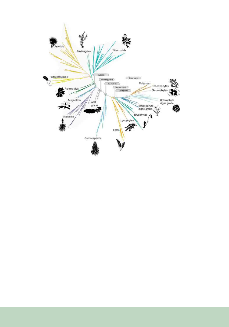
196
Discussion
Scientific figures can quickly and concisely
communicate detailed information, but without
clear visual landmarks, they can be impenetrable.
In biology, silhouettes are an easy way to instantly
convey taxa to other scientists and the public.
PhyloPic’s phylogenetic organization and open API
make it simple to find and use relevant silhouettes
for a given taxon. All silhouettes on PhyloPic are
hosted under Creative Commons licenses in the
public domain (PDM), CC0 (requires no credit
given), or as CC BY (requires credit given to the
author). It is up to the end-user to properly credit
silhouette authors and PhyloPic when necessary.
To assist with the creation of silhouettes, PhyloPic
has a “Silhouette Sponsorship” program (https://
phylopic.org/sponsorship), where users can
commit to creating silhouettes for their chosen
taxon or donate to PhyloPic to request taxa to be
added. Requested taxa are placed on a publicly
accessible priority list that allows volunteer
contributors to prioritize silhouette creation.
However, creating a silhouette of your chosen
organism following our how-to videos (Figure 1)
is the most efficient option.
PhyloPic affords plant scientists the opportunity
to use free silhouettes on statistical charts,
biogeographic maps, cladograms, and other data
visualizations. However, plants are currently
under-represented in PhyloPic, limiting the
potential of this community-led tool. For
perspective, if every member of the Botanical
Society of America (n ≈ 3000, 2023) contributed
just one silhouette, there would be nearly five
times as many available plant silhouettes. With the
Figure 2. A sample figure adapted from “One thousand plant transcriptomes and the phylogenomics
of green plants” (Leebens-Mack et al., 2019) with silhouettes from PhyloPic added. Silhouettes down-
loaded from PhyloPic: https://www.phylopic.org/collections/a7844e00-c254-07de-0137-ccb687820a6d

197
submission process recently re-engineered and
streamlined, we encourage everyone to contribute
to this valuable community resource.
AUTHOR CONTRIBUTIONS
M.M.: Conceptualization, Methodology, Data
Curation, Writing, Visualization, Supervision,
Project Administration; M.K. Conceptualization,
Methodology, Validation, Resources, Writing
DATA AVAILABILITY
STATEMENT
All how-to videos and data used in this paper are
freely available on the Open Science Framework
repository (doi.org/10.17605/OSF.IO/JBUW5),
on PhyloPic.org, or by request from the
corresponding author.
REFERENCES
Adobe Inc. 2023a. Adobe Illustrator. Website:
https://
adobe.com/products/illustrator
Adobe Inc. 2023b. Adobe Photoshop. Website:
https://
www.adobe.com/products/photoshop.html
Berg, S., D. Kutra, T. Kroeger, C. N. Straehle, B. X. Kausler,
C. Haubold, M. Schiegg, et al. 2019. ilastik: interactive ma-
chine learning for (bio)image analysis. Nature Methods 16:
1226–1232.
Gearty, W., L. A. Jones, and S. Chamberlain. 2023. rphy-
lopic: an R package for accessing and plotting PhyloPic sil-
houettes. TBD.
Inkscape Project. 2023. Inkscape. Website: https://inkscape.org
Kirillov, A., E. Mintun, N. Ravi, H. Mao, C. Rolland, L. Gus-
tafson, T. Xiao, et al. 2023. Segment Anything. ArXiv Web-
site: https://arxiv.org/abs/2304.02643.
Leebens-Mack, J. H., M. S. Barker, E. J. Carpenter, M. K.
Deyholos, M. A. Gitzendanner, S. W. Graham, I. Grosse, et
al. 2019. One thousand plant transcriptomes and the phy-
logenomics of green plants. Nature 574: 679–685.
McClennen, M., J. Jenkins, and M. Uhen. 2017. The Paleo-
biology Database.
OpenTreeofLife, K. A. Cranston, B. Redelings, L. L. S. Reyes,
J. Allman, E. J. McTavish, and M. T. Holder. 2019. Open
Tree of Life Taxonomy. Website: https://doi.org/10.5281/ZE-
NODO.3937751
Rueden, C. T., J. Schindelin, M. C. Hiner, B. E. DeZonia, A.
E. Walter, E. T. Arena, and K. W. Eliceiri. 2017. ImageJ2:
ImageJ for the next generation of scientific image data. BMC
Bioinformatics 18: 529.
Schindelin, J., I. Arganda-Carreras, E. Frise, V. Kaynig, M.
Longair, T. Pietzsch, S. Preibisch, et al. 2012. Fiji: an open-
source platform for biological-image analysis. Nature Meth-
ods 9: 676–682.
Schneider, C. A., W. S. Rasband, and K. W. Eliceiri. 2012.
NIH Image to ImageJ: 25 years of image analysis. Nature
Methods 9: 671–675.

198
From the P SB Special I ssue on Art in the Botanical Sciences
Humans transform, filter, break down, and
reassemble seemingly endless amounts of
information as we make sense of the world we
live in. For an ecologist observing a natural
ecosystem, this process may produce a graphical
figure summarizing a targeted property of the
system to understand the consequences of
environmental change; for an artist observing the
same phenomenon, this may result in the formal
use of abstraction, form, and color as an inquiry
into the ways humans interface with “Nature.” If
viewed only in this way. the two disciplines emerge
as a simple dichotomy; yet in reality, the two have
many approaches in common, both of which help
us understand the world we live in. Since 2014, the
two of us (a scientist who studies the ecological
interactions among of plants and animals, and a
visual artist utilizing video and digital media) have
been collaborating at the intellectual confluence of
the sciences and arts with the goal of generating
novel perspectives on the world that surrounds
us and our relationships to it. In this paper, we
Can the Collaboration of Science and Art
Broaden Our Understanding of Nature?
Paul J. CaraDonna
1,2,3,4
Mark Dorf
1,4c
1
Rocky Mountain Biological Laboratory, Crested Butte,
CO, USA
2
Chicago Botanic Garden, Glencoe, IL, USA
3
Plant Biology & Conservation, Northwestern University,
Evanston, IL, USA
4
Correspondence: pcaradonna@chicagobotanic.org and
mdorf@mdorf.com
discuss our long-term, ongoing collaborations
at the intersection of science and art, how it
can influence our individual perspectives by
building trust and exchange between scientists
and artists, and how such collaborations have
the potential to create new ways of knowing and
understanding. To explore these ideas, together
we created Figure 1: a creative appropriation
of video still’s taken from Dorf’s (2021) film, A
New Nature, that was predominantly produced
and conceptualized during the 2021 field season
at the Rocky Mountain Biological Laboratory.
Throughout the film, the viewer is provoked to
consider not only the future of what Western
culture commonly refers to as “Nature” in the face
of a changing planet, but also what the term itself
means in contemporary life and language. Figure
1 functions as an illustration of the work produced
from our long-standing collaboration, as well as a
visual tool and metaphor to better understand the
ways in which we collaborate.
Ways of Seeing Nature
Taking influence from John Berger’s Ways of Seeing
(1972), we begin by asking the question: when
observing nature, what is it that we see and why
do we see it in that way? This question sits at the
center of our collaboration as scientist and artist
and is one that we are continually investigating.
Figure 1A presents an image that can feel both
familiar and foreign. Recognizable elements of a
spruce forest can be identified on a localized scale:
needle-like leaves, tree trunks, color palette, and
variation in light. But when zooming out to see
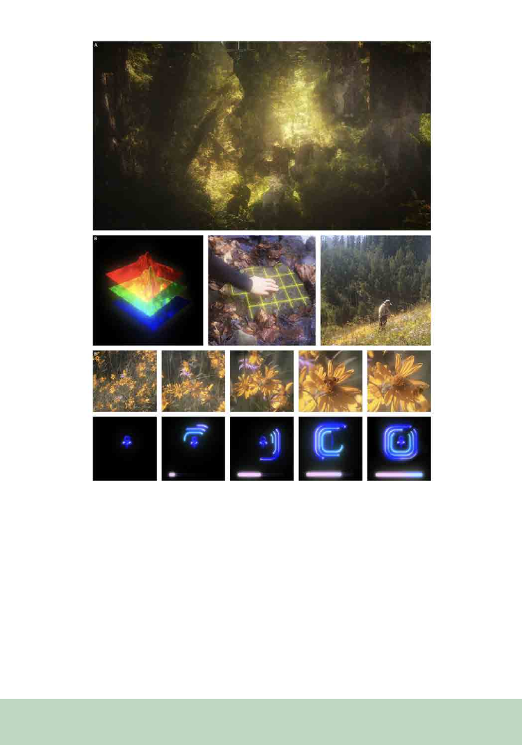
199
the entire image, these familiar elements and the
logic of the image and the forest itself fall apart. We
have something that appears and feels like a forest,
while simultaneously approaching nonsense. In
other words, Figure 1A asks us to ponder: how can
we recognize this image of a forest when there is
in fact no forest represented; or, put another way,
how do we understand something as it changes
rapidly in real time? This idea is analogous to
how, as we begin to understand many dimensions
of ecological systems for the first time, they are
simultaneously shifting in response to global
change.
Figure 1. An illustration of the work produced from the bidirectional collaboration between science and art as a visual tool and
metaphor of the collaboration and how we understand nature. We created this figure together in Dorf’s art studio in New York
City and then later at CaraDonna’s lab at a remote biological field station in Colorado (The Rocky Mountain Biological Labora-
tory). (A) Digital collage image of subalpine spruce forests. (B) Digital image of three identical 3D-rendered mountains split into
the additive color channels of red, green, and blue. (C) Digital image of a hand reaching into a small pool of water that has a
grid atop its surface. (D) A scientist in a subalpine meadow noting the timing of an observation of a pollinator visiting a plant.
(E) Two series of five images illustrating the passing of time building up to a discrete event. All still images are from the film, A
New Nature (Dorf, 2021).

200
As we consider this tension in our observation
and perception of nature, we turn to Figure 1B,
which investigates how we filter, transform, split,
and rearrange our observations as we try and
make sense of them. In Dorf’s (2021) film, A New
Nature, the form featured in Figure 1B rotates,
undulates, and slowly separates as the voiceover
asks: “Tell me what you see. Can you tell me
what I see? What is there in front of you?” The
voice over and rotating landscape are directly
inspired by Piaget’s (1954) experiments with early
childhood development and object permanence
in which Piaget would present a model landscape
to a child, ask them to describe the scene, turn the
landscape, and continue to ask probing questions
to see if the child recognized the landscape as
the same form. The constant state of fluctuation
renders Piaget’s request to describe the subject’s
state nearly impossible. In the case of A New
Nature (Dorf, 2021), the viewer is presented with
an impermanent moving target that is not only in
constant rotation, but also changing form from
one moment to the next. Eventually the rotating
landscape divides into three identical 3D-rendered
mountains split into the additive color channels
of red, green, and blue. When separated they
are independent entities, but when combined
they compose an image that represents the fully
realized spectrum of color and light. Functionally,
digital images are presented with all their color
channels combined together so that the image is
more or less a reflection of a sensory experience
or an observation; similarly, scientific ideas are
presented as cohesive and generalized frameworks
that otherwise emerge from many disparate
sources of empirical information. When an image
is broken apart, as in Figure 1B, the viewer is
challenged to consider how even the most basic
ideas, observations, and environments can be
infinitely split or combined, revealing something
foreign and strange in a new and unexpected
manner. This is a process that both scientists and
artists are constantly enacting.
Conduits for Collaborative Seeing
The question of asking what one sees and how
one sees it is a more complicated inquiry than
it might seem. In the case of the 3D-rendered
mountain splitting apart (Figure 1B), the challenge
presented is that if everything is in constant flux,
how do we interpret what we are observing?
Despite their different approaches, scientists and
artists both run up against this problem, whether
considering, for example, plants adapting to
rapidly changing climate conditions, or the ever-
shifting relationship among humans, technology,
and nature. Figure 1C and 1D illustrate two
different moments of interfacing with the world.
Figure 1C shows the hand of an artist reaching
into a small pool of water only to be met with a
graphic grid that lays atop the water itself; Figure
1D shows a scientist in the field noting the timing
of an observation of an interaction between plant
and pollinator. What both images help to reveal is
that the human observer is the conduit for seeing,
observing, and understanding. The scientist and
the artist bring with them different histories and
toolsets for interfacing with the world, but they
both share the common feature of the human
acting as the filter for translating the sensory
experience of the world—the observation—into
knowledge.
The methods of observation of the scientist and
artist can be very different, but something that
is not so obvious is that the goals of the scientist
and artist are often shared. Figure 1E investigates
this idea with two series of images that illustrate
a narrative arc of the passing of time building up
to a discrete event. The top row illustrates a slow
zoom sequence of the formation and dissolution
of an interaction between a flower and a bumble
bee; the bottom row illustrates the sequence
of a graphic-loading interface of a unicode
flower, which spins in the center as the sequence
progresses. From one perspective, they represent
two divergent narrative arcs as seen from their

201
deeply contrasting aesthetic representations. But
from another perspective, they both center on the
passage of time and the temporal sequence of the
completion of a natural event (e.g., CaraDonna
et al., 2014, 2017, 2021; Post, 2019). What Figure
1E seeks to interrogate with the juxtaposition of
these two sequences is the question of seeing,
observing, and interpretation. As with the other
elements of Figure 1, we continually ask: what are
we seeing, who is doing the seeing, what is the
mode of observation, and how is it all interpreted?
Critically, it is not so much that one way of seeing
or knowing is better or more accurate—instead, we
argue that together, we have a fuller understanding
of the world that reaches beyond that of the
quantitative, qualitative, logical, and emotional
.
Novel Ways of Knowing Nature
The scientist and the artist can begin with the same
source of influence and the same set of information
(plants, animals, and their interactions); use
different means of analyzing, observing, and
understanding (population dynamics, ecological
networks; color, light, sound); and naturally resolve
on quite different results (e.g., CaraDonna et al.,
2017; Dorf, 2021). If the collaboration between the
two is simply art in service to science, or science
in service to art, then new ways of knowing do not
easily emerge. What we have learned over nearly
10 years of bidirectional sharing at the confluence
of science and art is that there is much to be gained
through trustful and open collaboration. Such
collaboration has helped to reveal to us that there is
no one result that is greater than the other. Instead,
we find that knowing and knowledge production
is constantly in flux like the surroundings that
seed our inquiries. If we allow it, each approach
informs the other, helps to challenge their mutual
assumptions, and shifts perspectives.
ACKNOWLEDGMENTS
We thank Amy Iler, Cassandra Croft, Cory
Zimmerman, Nick Waser, Mary Price, Jane
Ogilvie, Will Petry, and the CaraDonna Lab for
stimulating discussions and support. We thank two
anonymous reviewers for their insight comments
on the manuscript. We thank the Rocky Mountain
Biological Laboratory (RMBL) for providing
access to field sites and logistical support. Funding
was provided by the RMBL, Northwestern
University, and the National Science Foundation
(DGE-1754518 to P.J.C.).
REFERENCES
Berger, J. 1972. Ways of Seeing. British Broadcasting Com-
pany.
CaraDonna, P. J., A. M. Iler, and D. W. Inouye. 2014. Shifts
in flowering phenology reshape a subalpine plant commu-
nity. Proceeding of the National Academy of Sciences, USA
111: 4916–4921.
CaraDonna, P. J., W. K. Petry, R. M. Brennan, J. L. Cunning-
ham, J. L. Bronstein, N. M. Waser, and N. J. Sanders. 2017.
Interaction rewiring and the rapid turnover of plant–pollina-
tor networks. Ecology Letters 20: 385-394.
CaraDonna, P. J., L. A. Burkle, B. Schwarz, J. Resasco, T. M.
Knight, G. Benadi, N. Blüthgen, et al. 2021. Seeing through
the static: the temporal dimension of plant–animal mutualis-
tic interactions. Ecology Letters 24: 149-161.
Dorf, M. (Director). 2021. A New Nature. [Film]
Piaget, J. 1954. The development of object concept (M.
Cook, Trans.). In: J. Piaget, M. Cook (Trans.), The construc-
tion of reality in the child (pp. 3–96). Basic Books.
Post, E. 2019. Time in Ecology: A Theoretical Framework
[MPB 61], Princeton: Princeton University Press.

202
From the P SB Special I ssue on Art in the Botanical Sciences
The combination of art and the botanical sciences
can address global and local issues in ways
that resonate with communities. Marginalized
societies, plant species, and whole ecosystems
are suffering the consequences of a rapidly
changing climate (IPCC, 2023). These challenges
require multidisciplinary approaches, which
extend beyond the field of botanical science. We,
the authors, propose that socially engaged art
practices rooted in a history of activism combined
with a deep western scientific understanding of
plant communities can help address the issues
of climate change. We are a group of creative
researchers from various disciplines, all part of
the Backyard Phenology Project (BYP) at the
The Integration of Botanical Science,
Art, and Agency
Maria Park
1,7
Jessie Merriam2
Abbie Anderson3
Chotsani Elaine Dean4
Rebecca Montgomery5
Christine Baeumler6
1
University of Minnesota (UMN) PhD candidate in
Ecology, Evolution, and Behavior; artist
2
UMN Masters in Heritage Studies and Public His-
tory; artist
3
UMN Researcher, Department of Forest Resources
4
UMN Assistant Professor of Ceramics, Department
of Art
5
UMN Professor of Forest Ecology, Department of
Forest Resources
6
UMN Professor of Interdisciplinary Art & Social
Practice, Art Department Chair
7
Author for correspondence: mariap@umn.edu
University of Minnesota (UMN). In this article,
we describe this project and related endeavors.
We hope that these examples can help readers see
their place in both local and global ecosystems
and build communities through the integration of
the arts and botanical sciences.
Backyard Phenology
The BYP is an ongoing art and community
science platform that engages people to become
more attuned to the seasonal cycles of nature
(phenology) and foster dialogue about the
influence of climate change on nature (Backyard
Phenology, 2023). The project launched in 2016,
with support from the Northern Lights arts
organization and a UMN Grand Challenges
grant. The project was founded by Professor
Rebecca Montgomery, a forest ecologist, and
Professor Christine Baeumler, a socially engaged
artist. Other founding collaborators include
Professor Mae Davenport, Beth Mercer-Taylor,
and Kate Flick. The BYP team, assisted by various
researchers and volunteers, listen to and record
stories of people around Minnesota in a mobile
recording studio called the Climate Chaser.
Participants are invited to share stories of plants,
the environment, and our changing climate with
the BYP team, and then continue to document
their natural observations on a printed phenology
calendar. A BYP podcast featuring the recorded
stories is available on the BYP website https://
phenology.umn.edu. The Climate Chaser is a
retrofitted vintage chrome camper that has been
brought to numerous events and sites, including
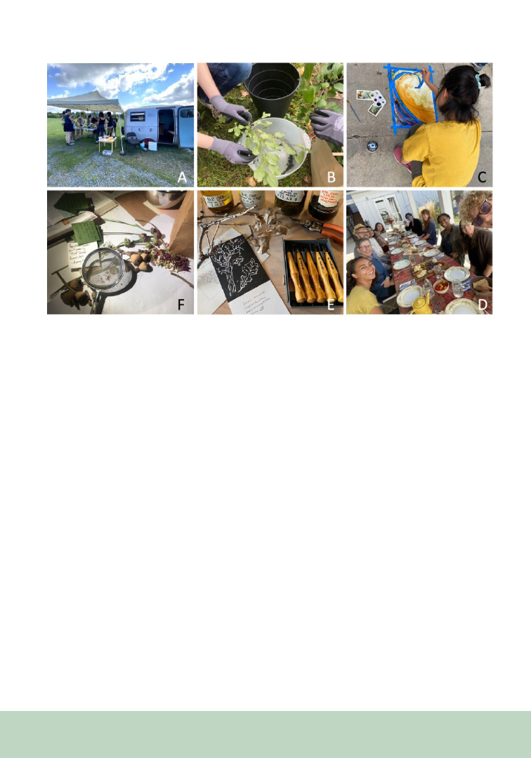
203
art festivals, an American Indian reservation, a
local farm, and the Minnesota State Fair. In 2022,
the camper visited Franconia Sculpture Park where
Baeumler invited Minnesota artist Kimberly
Boustead to lead a natural ink workshop alongside
the Climate Chaser (Figure 1A). BYP encourages
scientific observation and welcomes diverse ways
of knowing and experiencing the environment
through social engagement, collaboration, and
empowerment of participants.
Pedagogy
The themes and values of the Backyard Phenology
Project are fully present in the courses that
members of the BYP team co-teach at UMN.
The Art and Ecology course, currently taught by
Christine Baeumler and Maria Park, an artist and
graduate student in ecology, takes an experiential
and place-based approach to engaging students
in learning about ecology, eco-art, and socially
engaged art practices. Following the collaborative
BYP event at Franconia, artist Kimberly Boustead
joined Baeumler and Park to lead students in
harvesting common buckthorn berries (Rhamnus
cathartica) lining the Mississippi River. The
non-native species has been reported in every
Minnesota county and negatively impacts the
environment in many ways (EDDMapS, 2023).
Employees from the Minneapolis Park and
Recreation Board supplied tools and taught
students about the ecological implications of
Figure 1. (A) The Backyard Phenology Project: The Climate Chaser recording studio is parked next to a natural ink work-
shop tent hosted by Kimberly Boustead at Franconia Sculpture Park. (B) Pedagogy: Students in the Art and Ecology class
harvest buckthorn berries. (C) Maria Park and students experiment with shifting the pH of natural inks, thereby changing
the colors. (D) Reframing Our Relations: The multi-course meal was prepared by team members from foraged and locally
sourced ingredients including staghorn sumac, spruce tips, and crayfish. (E) A Plant People Project: bur oak (Quercus mac-
rocarpa) print and haiku next to various tinctures from oak bark. The class learned about the role of oaks in the Stop the
Reroute fight (1999), and its role in oak savannas. Check out the Plant People archive-in-progress: z.umn.edu/PlantPeople).
(F) Seed Sharing: Seed packet preparation by Chotsani Elaine Dean.

204
buckthorn. Boustead then led students through
the process of creating ink and painting with the
buckthorn berries (Figure 1B, C). In one class
period, students learned about the ecology, art,
and chemistry of a plant in a completely hands-on
experience.
During the semester, students in Art and Ecology
find a “sit-spot” location that they return to
consistently. Based on the practice of taking
phenology measurements, students are prompted
to observe changes in the environment and reflect
on their chosen location as the seasons shift.
Setting aside the time to regularly disconnect from
the busyness of everyday life and connect with a
place in nature has helped students care for their
mental health in addition to the environment. The
sit-spot practice is also fostered in a UMN Grand
Challenges Curriculum course titled Making
Sense of Climate Change: Science, Art, and Agency,
currently taught by Montgomery, Baeumler, and
Par
k.
Reframing Our Relations
As noted above, Franconia Sculpture Park has
been a site of artistic and ecological engagement
for the BYP team. In 2022, the BYP team, led
by BYP member Christian Bell, hosted an event
titled, “Re-framing our Relations: Complicating
the invasive species narrative through participatory
art and science at Franconia Sculpture Park.” The
interdisciplinary team of artists and scientists
engaged local stakeholders and community
members in the sharing of food, stories, and
knowledge about non-native species (Figure
1D). A major goal of the project was to reframe
the ways in which introduced species are viewed
and managed. A more respectful vocabulary of
animacy (Kimmerer, 2017) could be used instead
of colonial language that vilifies species by referring
to them as “invasive,” “novel weapons,” and in need
of “eradication” (Estévez et al., 2015). Learning
from Indigenous practices of land stewardship,
and opening minds to the possibilities of long-
term ecological restoration practices, can allow
for a more sustainable future (GLIFWC Climate
Change Team, 2023).
The act of facilitating conversations over a shared
meal is a socially engaged art practice that has
been implemented by contemporary artists such
as Seitu Jones and Marina Zurkow (Gruenewald,
2014; Cadieux et al., 2019). This particular shared
meal and conversation was curated to encourage
participants to explore their own relationships
with plants, particularly non-native species.
Conversations spanned topics of ecology, land
management, colonialism, reciprocity, and
different ways of knowing. Participants exchanged
stories and ideated pathways forward together.
Plant People
Plant People is a research initiative documenting
oral histories of plant practitioners in Minnesota
to uplift the history of healing plant knowledge
in the midst of the Western medical tradition.
Jessie Merriam, a graduate research assistant
for Backyard Phenology and Plant People, was
inspired by BYP workshops at Franconia to help
Plant People identify the healing relationships
artists also have with plants. Merriam and
herbalist Emily Ryan developed an ongoing series,
“The Ways of Plants,” at Fireweed Community
Woodshop in Minneapolis to explore the history,
sustainable harvest, and culinary and medicinal
uses of native plants while teaching students to
make their own woodblock print of the plant
(Figure 1E).
Seed Sharing
Seed harvesting and sharing is a project that is
essential in the studio practice of Chotsani Elaine
Dean, a member of BYP and Assistant Professor
in the UMN Art Department. While living in
South Carolina in 2017, Dean learned where to
find the seeds of a flowering plant by spending
time with two of her garden plants. When Dean

205
noticed the open and offering seed pod of the
pansy (Viola × wittrockiana) and nasturtium
(Tropaeolum majus), her curiosity was sparked.
Dean learned how to identify the flowers, followed
the phenology of the plants, wrapped flowerheads
in cheesecloth to catch seeds, and started to make
her own seed packets (Figure 1F). Dean continues
to give people her seed packets to continue the
practiced tradition of across-the-fence gardening
in this modern era.
Conclusions
The intersections of art and the botanical sciences
can foster innovative community engagement
initiatives that address the pressing challenges
of climate change and dominating non-native
plant species. People may initially question and
misunderstand the role of socially engaged art
in the botanical sciences. But by helping people
interact and reflect upon plants and natural places
they care about, we have seen shifts in attitude
and perspective. We have found that approaching
projects with an open mindset and willingness to
engage with people from different backgrounds
lead to exciting collaborations that cross societal
boundaries and generate opportunities to co-
create a sustainable world.
ACKNOWLEDGMENTS
We are thankful for everyone who has been
involved with the projects, including team
member Christian Bell, collaborating artist
Kimberly Boustead, past BYP coordinators Sam
Graf and Jonelle Walker, researchers Emily Green
and Bree Duever, UMN graduate students Jessica
Lackey and Jenn Shepard, UMN undergraduates
Rachel Nichols, K. Arsten Lennartson, and
Lauren Schultz, and Professor Nicholas Jordan,
an original founding member of BYP. We are
grateful for funding from Northern Lights and
former artistic director Steve Dietz, the University
of Minnesota Grand Challenges Research Award,
and the Institute on the Environment at UMN
Mini Grant. The Climate Chaser camper rehab
was done by ThreeSeven, graphics were created by
This is Folly, and printing of BYP calendars were
done by Smart Set.
REFERENCES
Backyard Phenology. 2023. The University of Minnesota.
Website: https://phenology.umn.edu/.
Cadieux, K. V., M. Zurkow, and S. Libertus. 2019. Eating
Climate Change with Making the Best of It Dandelion: Col-
laborative Public Art as a Mode of Future-Oriented Learning.
Transformations: The Journal of Inclusive Scholarship and
Pedagogy 29: 210–233.
EDDMapS (Early Detection & Distribution Mapping Sys-
tem). 2023. The University of Georgia - Center for Invasive
Species and Ecosystem Health. Website: http://www.edd-
maps.org/.
Estévez, R. A., C. B. Anderson, J. C. Pizarro, and M. A.
Burgman. 2015. Clarifying values, risk perceptions, and at-
titudes to resolve or avoid social conflicts in invasive species
management: Confronting Invasive Species Conflicts. Con-
servation Biology 29: 19–30.
GLIFWC Climate Change Team. 2023. Aanji-bimaadi-
ziimagak o’ow aki. Great Lakes Indian Fish and Wildlife
Commission, Odanah, WI. 332 pp.
Gruenewald, B. 2014. A recipe for change: Public Art St.
Paul to host community meal for 2,000 guests. Hmong Times
September 3, 2014: 3.
IPCC, 2023: Climate Change 2023: Synthesis Report. Con-
tribution of Working Groups I, II and III to the Sixth As-
sessment Report of the Intergovernmental Panel on Climate
Change [Core Writing Team, H. Lee and J. Romero (eds.)].
IPCC, Geneva, Switzerland, pp. 35-115, doi: 10.59327/
IPCC/AR6-9789291691647.
Kimmerer, R. W. 2017. Learning the Grammar of Animacy.
Anthropology of Consciousness 28: 128–134.
206
From the P SB Special I ssue on Art in the Botanical Sciences
Since 2003, Cornell University’s Art of
Horticulture course has engaged students to
use plants for creating art (natural dyes, willow
weaving, papermaking, etc.) as well as the subject
of art (botanical illustration, photography,
printmaking, etc.). But founder Marcia Eames-
Sheavly’s learning objectives went far beyond
the basics of plants and paints, challenging the
traditional boundaries of university horticulture
curricula. This unique course encourages students
to develop a distinctive lens to view the world and
to take a deep exploration into self, the classroom
environment, and their broader community.
The curriculum has continued to evolve and
respond to changes in our scientific knowledge
and social awareness under the leadership of
“Art of Horticulture” Course
Cultivates Creativity
Emily Detrick
1
Craig Cramer
2
1
Director of Horticulture, Cornell Botanic Gardens and
Lecturer, School of Integrative Plant Science (SIPS),
Cornell University
2
Communications Specialist, School of Integrative Plant
Science (SIPS), Cornell University
Emily Detrick, who has taught the course since
2018.
The course vision is as follows:
Ground Students in the Landscape
Detrick has strengthened deep connections
between the course and Cornell Botanic Gardens,
where she serves as Director of Horticulture.
Plants from the Botanic Gardens provide both
inspiration and materials for creating art and a
seasonally changing palette that grounds students
in the cycles of the natural world. The gardens
offer a living laboratory where students participate
in activities like a mindful botany walk, create
plant-based installations that are accessioned into
the living collections, and learn about biocultural
diversity through interpreted displays (Figure 1).
Cultivate a Diverse and Inclusive
Classroom Culture
Although based in Cornell’s College of Agriculture
and Life Sciences, students come from across
disciplines and with varying experiences in
horticulture and art—majors as diverse as
Engineering, Animal Science, Computer Science,
Hotel Administration, and Architecture, in
addition to Plant Sciences and Fine Art. The
weekly sessions help students connect creativity
and botanical sciences through hands-on activities
that hone observation and plant biology skills as
well as an understanding of the principles of design
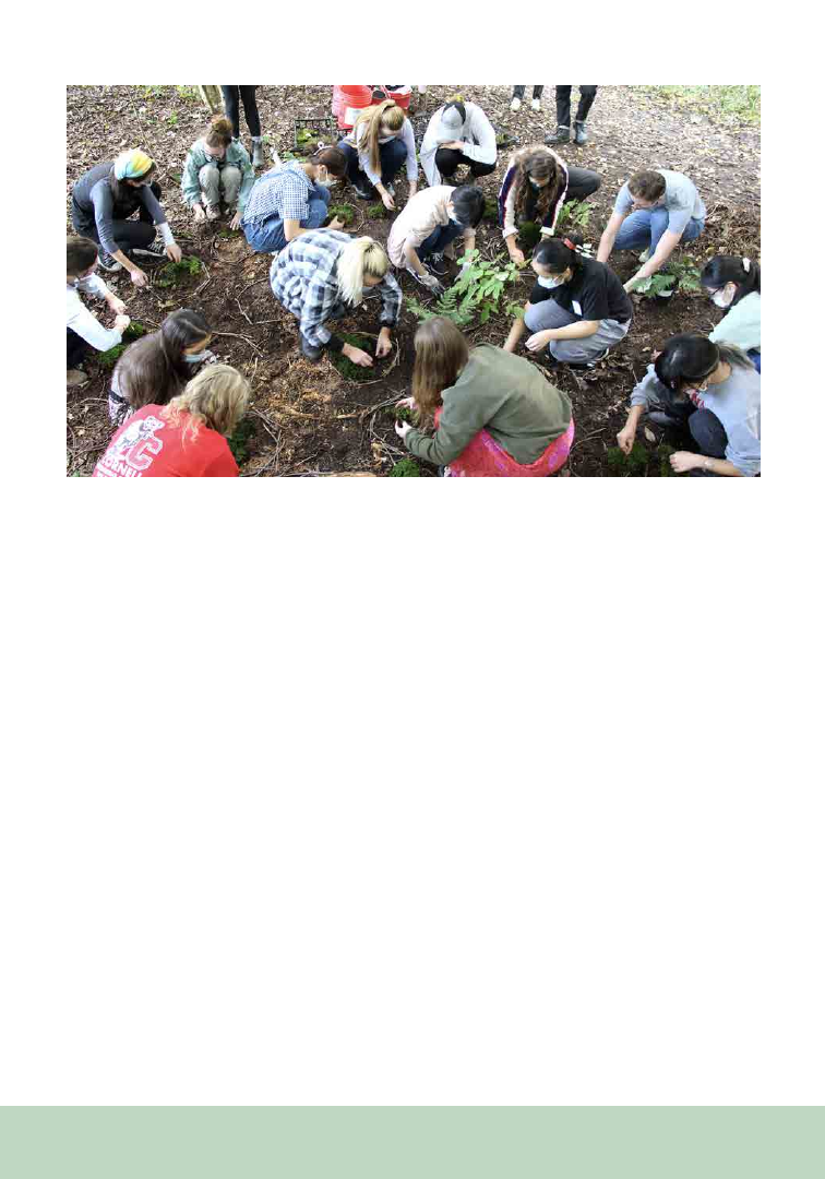
207
in living forms. Students weave their personal
experiences and interests with their botanical
arts exploration, a process that naturally creates
vulnerability. They engage with their peers to ask
questions, seek ideas, and support one another
when stepping out of their comfort zones. These
practices help develop group as well as individual
problem-solving strategies and contribute to an
inclusive, collaborative classroom culture.
Connect to Art-Science Community
Perhaps as important as the curriculum is the
insight students gain into a variety of career
paths through interaction with contemporary
artist-scientists. In addition to hosting artists in
the classroom, students take field trips to a local
gourd artist’s studio and a fiber artist’s dye garden.
Additionally, students engage with renowned
artists who visit campus, such as Brazilian street
artist Eder Muniz, who created a two-story
botanical mural at the Botanic Gardens in 2019.
Nigerian-American artist and poet Precious
Okoyomon led students in a nature-inspired
poetry workshop in 2022.
Deepen Knowledge of Campus
Programs and People
Students visit Cornell University’s Herbert F.
Johnson Museum of Art and Cornell Rare and
Manuscript Collection to see examples of historic
and contemporary plant-inspired art. Cornell
artists-scientists lead classes, such as Cornell Soil
Health Lab manager Kirsten Kurtz, who teaches
the importance of soil health as well as how to
paint with it. SIPS Communications Specialist
Craig Cramer teaches botanical scanning and
digital manipulation. He also documents student
work and manages the course website, where
images of past student work can be found (https://
blogs.cornell.edu/artofhort/).
Figure 1. Art of Horticulture students create moss “land art” installation at Cornell Botanic Gardens using native mosses
and ferns. If conditions are right, the students’ individual moss arrangements will grow and merge into a single, natural-
ized composition of brocade moss (Hypnum imponens), haircap moss (Polytrichum commune), and marginal wood fern
(Dryopteris marginalis).

208
Develop Observation Reflection and
Documentation Skills
Students learn creative ways to document their
observations of plant life, such as cyanotypes,
pressing and drying techniques, and ink
impressions. Required readings and writing
assignments hone their reflection skills to connect
self, art, and the natural world in a broader
context. Students apply their unique interests,
perspectives, and expertise to create capstone
projects. When presenting, they show evidence of
their new botanical understandings by discussing
the nomenclature, morphology, life histories, and
ethnobotanical uses of plants they have come to
know intimately through their projects, as well as
their successes and challenges with newly learned
artmaking techniques.
Create Lifetime Impacts
Overall, students learn the value of engaging
creatively with plants to foster their own and
community well-being. One reflected, “I have
gained… lessons not only about the intersection
of art and science, but about reflecting in myself,
taking time to look up at the natural world around
me, connecting with my peers, using my voice
with intention, and trying things that scare me.”

209
From the P SB Special I ssue on Art in the Botanical Sciences
With increasing anthropogenic and climate-
induced changes to our ecosystems (Groffman,
2014; Kidwell, 2015), it is more imperative than
ever to foster a kinship between people and their
environment and to enhance communication
of ecological knowledge across audiences, from
scientists to students and the public. Cultivating
a personal connection to plants—from individual
species to their foundational role in ecosystems—
is especially critical, because doing so promotes a
desire for conservation and a deeper understanding
of our world’s biodiversity. While many media
platforms, such as nature documentaries, tend to
focus on mammals (Howlett et al., 2023), plants
account for 80% of total biomass on Earth (Bar-
On et al., 2018) and serve as the building blocks of
food webs. Yet nearly 40% of all plant species are
classified as rare (Enquist et al., 2019).
Reaching Across Audiences:
Connecting to and Communicating
Botanical Concepts Through Art
Janette L. Davidson
1,5
Sienna A. Wessel
1,2
Jennifer W. Shoemaker
3
Alexandra P. Rose
4
Lauren G. Shoemaker
1
1
Botany Department, University of Wyoming, Laramie,
Wyoming 82071 USA
2
Colorado Forest Restoration Institute, Colorado State
University, Fort Collins, Colorado 80521 USA
3
Fine Artist, Fort Collins, Colorado 80526 USA
4
Institute of Arctic and Alpine Research, University of
Colorado, Boulder, Colorado 80303 US
A
5
Author for correspondencce: jdavid21@uwyo.
edu
Artwork presents one key avenue for promoting
botanical literacy by creating an emotional, cross-
scale connection to plants and fostering a bigger-
picture context of plant responses to climate
change. While detailed and highly accurate
botanical illustrations date back centuries and
were crucial in early efforts to record plant species
prior to photography, recent trends in ecological
education in STEAM (science, technology,
engineering, art, and math) have moved toward
a more holistic view of plants embedded within
their abiotic and biotic environments. Parallel
trends are occurring in the world of fine art, with
pieces focusing on fostering a personal connection
between humans and the botanical world. Here
we examine how art promotes this connection
between people and plants across biological scales.
Individual and Species-Level Scale
One of the most direct paths humans may
take to connecting with plants is by bringing
houseplants into their homes. Greening a home
through the introduction of houseplants benefits
mood and task attentivity, with even individual
plants inspiring creativity (Shibata and Suzuki,
2002) and motivating artistic compositions that
further botanical understanding. Houseplants
skyrocketed in popularity during the COVID-19
pandemic (Phillips and Schultz, 2021), quickly
becoming muses for creative projects such as
leaf preservations, still life paintings, and artfully
arranged terrariums. Plants also inspired artistic
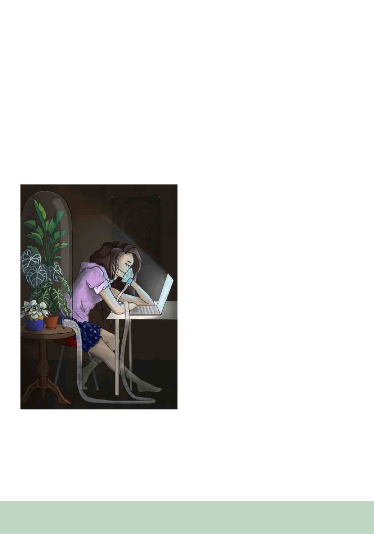
210
competitions, such as one hosted by the Denver
Botanic Gardens inviting artists to submit pieces
representing how houseplants, gardens, and
nature supported them through the COVID-19
pandemic (Denver Botanic Gardens, 2021)
(Figure 1). During this time, an increase in
emotional, sentimental attachment to plants
also became evident. In addition to inspiring
creativity, houseplants also benefit mental health
as well as encourage mindfulness and self-care
(Bringslimark et al., 2009). It is perhaps for this
reason that many people who were quarantined
away from both social and ecological interactions
came to see their houseplants as sources of
personal joy, inspiration, and comfort during this
time. Through art, this quiet connection may be
succinctly and poignantly shared with any who
may relate or sympathize with a similar connection
to their personal plants.
Interactions with plants at an individual and
personal scale also improved many owners’
knowledge of botany. Plant ownership exposes
non-academics to botanical concepts such
as environmental requirements, anatomical
terminology, key features for identification, and
conservation concerns such as poaching of rare
plants. From this increased knowledge arose
artistic infographics and care sheets, spreading
information on these concepts in a visually
accessible medium (Briscoe, 2020).
Community and
Ecosystem-Level Scale
The positive associations art can engender
with indoor plants may be extended to connect
people with larger natural systems, and botanical
illustration continues to play a valuable role in the
study of ecosystems. Artists may also look beyond
the individual to capture the lives of plants within
their larger landscape context.
Landscape fine artists often create artwork of
plant communities at this moderate scale, moving
beyond detailed renderings into the sphere of
artistic interpretation of plants within their
ecosystems. Using observation and knowledge
of plant communities, artists convey the sense
of a landscape using loose brushwork and rich
color. The popular plein air movement (a French
phrase meaning “in open air”), where artists are
simultaneously inspired by and create directly
in nature, began in the early 1800s, and was
popularized during the Impressionism Movement
(Callen, 2015) and continues to this day. This
technique captures the effects of sunlight and
shadow on a landscape, allowing artists to convey
their emotional connection and impressions of
Figure 1. Life Support, 2021. Digital art by Janette David-
son. Image symbolically depicts a connection between people
and plants, in how houseplants supported their caretakers
through the quarantine period of the COVID-19 pandemic.
This piece was created for the 2021 online art competition
hosted by the Denver Botanic Gardens, “Plants Through
Pandemic.”
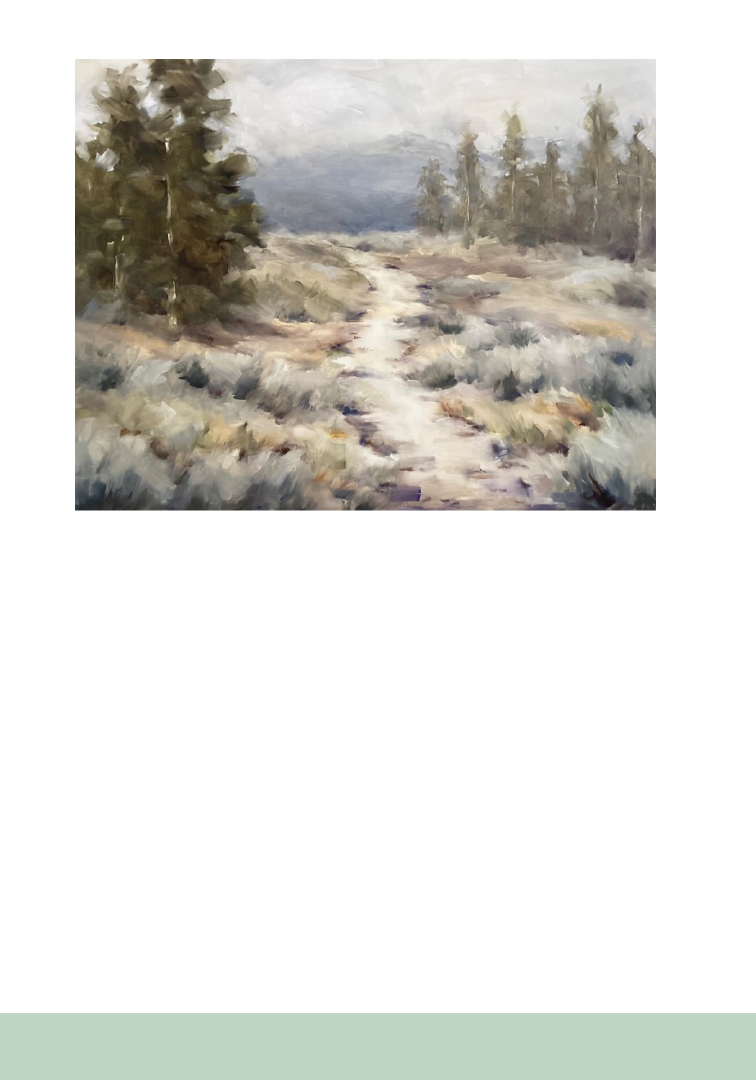
211
the landscape directly to viewers (The Art Story
Foundation, 2023) (Figure 2). Through landscape
art, viewers are invited to become absorbed
in the feel of a plant community, developing a
sense of place and appreciation at an ecosystem
level (Malafronte, 2009). In this medium, we
see ourselves not only observing the world of
plants within their landscapes, but becoming
immersed and ultimately belonging to that same
world. As such, these landscape paintings can
foster a personal desire to conserve ecological
communities (Ostendorf, 2017; Renowden et al.,
2022).
Global Scale
Art also has the potential to deepen general
understanding of abstract botanical concepts,
such as the risks of climate change to both humans
and plants. In the modern milieu, where 40% of
plant species face extinction due to threats such as
climate change (Antonelli et al., 2020), promoting
education of how plants are being affected on
a global scale is imperative in eliciting support
for conservation of these species. Through art,
seemingly abstract data such as changes in
temperature and climate patterns through time
(Figure 3) can come alive and inspire action.
However, communicating such topics, especially
to school-aged audiences, presents a challenge
because the concept may seem remote. An
interactive art medium is therefore well suited to
reach this audience and inspire students to take
action.
Figure 2. Perfect Day Ahead, 2023. Oil on panel by Jennifer Shoemaker. Landscape of sage and lodgepole pine
ecosystem, with a harmonious hiking trail snaking through. This piece was created as an example of integrating
historical and modern plein air techniques.
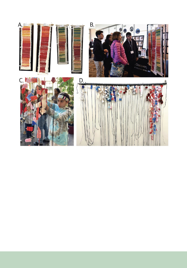
212
For young students, art can serve as a colorful
and captivating communicator of science—
particularly if the student is involved in the
creative process. Studies show that incorporating
art into learning environments may help students
better retain information (Gullatt, 2008), and thus
is a valuable tool in lessons about climate change.
Through the creative process, students can directly
connect to the data of their local environment
(Figure 3A, B), promoting understanding for how
this global problem can affect their smaller scale,
local botanical communities. A similar artistic
approach may be taken with communicating
global science to a multi-generational audience.
Involving citizens of varying ages can be powerful
for demonstrating trends through time at different
points in their lives, for example by asking
participants to attach color-coded cloth strips
representing the average temperature in a given
location during their birth year to a timeline
(Figure 3C, D). Having the public contribute
to interactive art exhibits at local community
events, such as at libraries or farmers’ markets,
can convey a concise visual message on climate
change impacts. Thereby, it can serve to initiate
conversations about climate change or encourage
families to contribute to conservation efforts
themselves by fostering an emotional connection
between plants, their ecosystems, and changes in
climate.
Figure 3. Collection of Tempestry (Barber and Gilson, 2023) educational and community artwork projects led by Dr. Al-
exandra Rose, depicting changes in temperatures over time in two North American cities: Mazatlán, Mexico and Boulder,
CO, USA (A, B), and illustrating the global temperature during the birth year of project participants at local community
events (C, D)
.

213
Promoting Scientific Literacy
through Emotional Engagement
Art’s distinctive ability to establish emotional
connections between viewers and the botanical
world across scales, spanning from individual
plants to botanical communities and a global
context, make it an effective tool for communicating
complex ecological concepts, eliciting pro-
environmental attitudes and behaviors in
society (Kals et al., 1999) and garner support
for solutions-focused conservation research. In
an era of global change, public understanding,
socio-political interest, and economic support
for plant conservation are more critical than ever
(Balding and Williams, 2016). However, people
often struggle to connect emotionally with plants,
and many of the concepts behind cutting-edge
conservation research can be difficult to convey to
non-specialists due to their abstract, multifaceted
nature. Education about conservation topics can
greatly enhance community engagement with
conservation work (Ardoin et al., 2020) and
scientists are increasingly reaping the benefits of
artistic approaches to communicating complex
topics (Curtis et al., 2012). Furthermore, art-
science integration can increase access to scientific
knowledge that can be restricted behind paywalls.
For example, plant functional traits are grounded
in theoretical concepts that can be used to generate
general predictions for how species may respond
to their environment and global change. However,
few people are well-acquainted with traits, and
the definition can be difficult to comprehend,
especially for mathematically derived botanical
traits such as specific leaf area (Violle et al.,
2007). In cases such as this, artwork can facilitate
understanding and engagement with botanical
concepts and conservation principles, such as
how traits are used by research scientists to predict
plant survival and adaptation in response to global
change (Figure 4). The brevity and illustrative
nature of the graphics served to share examples
of conservation work and increase social capital
for trait research by generating empathy for the
pressures that plants face. This approach is merely
one way art can distill abstract concepts into
engaging visual stories that stimulate empathy
and support for conservation. Other artists have
leveraged similar approaches, such as comic
strips (Kozik, 2021), to disseminate conservation
science in a problem-solution framework. Artistic
displays have immense potential to make science
more approachable and to arouse curiosity and
empathy for the botanical world.
Conclusions
Although art may appear, at first glance, to play a
fading role in the academic world of botany, it is in
fact one of our strongest tools for communicating
science. Most people have an innate connection
to nature, and whether it is strongest on a smaller
personal scale with individual plants or a larger
scale with landscapes, this connection can be
utilized to increase awareness about plants and the
challenges they face on a global scale. Botanical
art can also serve as a gateway to awareness and
aesthetic appreciation of the plant environment
and its associated organisms, from microbes to
pollinators. Art is a unique and valuable tool we
must use to tap into humanity’s connection with
plants, to not only improve the efficiency and
clarity with which we communicate science, but
also stoke a passion for botany and conservation
outside of the academic world.
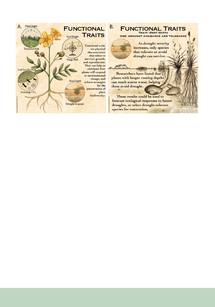
214
REFERENCES
Antonelli, A., C. Fry, R. J. Smith, M. S. J. Simmonds, P. J.
Kersey, H. W. Pritchard, M. S. Abbo, et al. 2020. State of
the World’s Plants and Fungi 2020. Royal Botanic Gardens,
Kew. https://doi.org/10.34885/172.
Ardoin, N. M., A. W. Bowers, and E. Gaillard. 2020. Envi-
ronmental education outcomes for conservation: A system-
atic review. Biological Conservation 241: 108224.
The Art Story Foundation. 2023. En Plein Air. Website:
https://www.theartstory.org/definition/en-plein-air/.
Balding, M., and K. J. H. Williams. 2016. Plant blindness and
the implications for plant conservation. Conservation Biol-
ogy 30: 1192–1199.
Barber, F., and J. Gilson. 2023. Stormy Weather: Textile art,
water and climate emergency. Textile 19: 1-13.
Bar-On, Y. M., R. Phillips, and R. Milo. 2018. The biomass
distribution on Earth. Proceedings of the National Academy
of Sciences 115: 6506-6511.
Bringslimark, T., T. Hartig, and G. G. Patil. 2009. The psy-
chological benefits of indoor plants: A critical review of the
experimental literature. Journal of Environmental Psychol-
ogy 29: 422-433.
Briscoe, P. 2020. Plant Care: Winter Is Coming. Reroot Gar-
dens 6 November 2020, Website: https://www.rerootgardens.
com/blogs/about/winter-is-coming.
Callen,
A. 2015. The Work of Art: Plein Air Painting and
Artistic Identity in Nineteenth-Century France. Reaktion
Books.
Curtis, D. J., N. Reid, and G. Ballard. 2012. Communicating
Ecology Through Art: What Scientists Think. Ecology and
Society 17(2).
Denver Botanic Gardens [@denverbotanic]. 24 Feb 2021.
Plants Through Pandemic Art Contest. Instagram. Website:
https://www.instagram.com/p/CLspg1Bj2Nb/
Enquist, B. J., X. Feng, B. Boyle, B. Maitner, E. A. Newman,
P. Møller Jørgensen, P. R. Roehrdanz, et al. 2019. The com-
monness of rarity: Global and future distribution of rarity
across land plants. Science Advances 5: eaaz0414.
Groffman, P. M., J. Cavender-Bares, N. D. Bettez, J. M.
Grove, S. J. Hall, J. B. Heffernan, S. E. Hobbie, et al. 2014.
Ecological homogenization of urban USA. Frontiers in Ecol-
ogy and the Environment 12: 74-81
Gulatt,
D. E. 2008. Enhancing student learning through arts
integration: Implications for the profession. The High School
Journal 91: 12-25.
Howlett, K., H.-Y. Lee, A. Jaffé, M. Lewis, E. C. Turner.
2023. Wildlife documentaries present a diverse, but biased,
portrayal of the natural world. People and Nature 5: 633-644.
Figure 4. Functional Traits, 2022. Digital art by Janette Davidson, authored by Sienna Wessel and Janette Davidson. This
shows two excerpts (A, B) from a five-part series of informational artistic graphics defining plant functional traits and
describing how they can be used to inform conservation efforts. The series was shared to a broad audience via university-
affiliated social media and a public gallery showing
.

215
Kals, E., D. Schumacher, and L. Montada. 1999). Emotional
Affinity toward Nature as a Motivational Basis to Protect Na-
ture. Environment and Behavior 31: 178–202.
Kidwell, S. M. 2015. Biology in the Anthropocene: Chal-
lenges and insights from young fossil records. Proceedings
of the National Academy of Sciences 112: 4922-4929.
Kozik, A. [@chase_prairie]. (2021, October 12). If you want
to read more about the differences between remnants and res-
torations…[Tweet]. Twitter. https://twitter.com/chase_prai-
rie/status/1447935165326635012/photo/1
Malafronte, A. 2009. The history of the plein air movement.
American Artist 73: 20-25.
Ostendorf, Y. 2017. Artists doing nature research. Website:
https://artistsandclimatechange.com/2017/03/16/artists-do-
ing-nature-research/.
Phillips, C., and E. Schulz. 2021. Greening home: caring for
plants indoors. Australian Geographer 52: 373-389.
Renowden, C., T. Beer, and L. Mata. 2022. Exploring inte-
grated ArtScience experiences to foster nature connectedness
through head, heart and hand. People and Nature 4: 519-533.
Shibata, S., and N. Suzuki. 2002. Effects of the foliage plant
on task performance and mood. Journal of Environmental
Psychology 22: 265-272.
Violle, C., M.-L. Navas, D. Vile, E. Kazakou, C. Fortunel,
I. Hummel, and E. Garnier. 2007. Let the concept of trait be
functional! Oikos 116: 882–892.

216
From the P SB Special I ssue on Art in the Botanical Sciences
“What is our mission? What do we have to do?
I think our mission is to reconcile, to reinte-
grate science and the arts, because right now
there’s a schism that exists in popular
culture. . .”
Mae Jemison, TED Talk (Jemison, 2002)
The schism between art and science is relatively
recent. Although scientific illustration has been
around for centuries, and many artists have
sought and seek inspiration from nature, after the
mid-19
th
century the disciplines dichotomized, as
scientists claimed their own space and the rise of
technology changed the world. In 1959, British
chemist, politician, and novelist C. P. Snow, in a
lecture titled "The Two Cultures and the Scientific
Revolution" (Snow, 1961), observed that the gulf
Lynne Gildensoph
Professor of Biology, Emerita,
St. Catherine University, St. Paul, MN 55105
Reconnecting Science with the Visual Arts:
Teaching the Art of Biology
between literary intellectuals and scientists was a
deterrent to human progress, since those in the
humanities had no way to communicate with
those in the sciences, and vice-versa.
Mae Jemison—first African-American female
NASA astronaut in space, MD, professor,
entrepreneur, and dancer—makes a powerful point
in her TED talk about this disconnect and argues
that it is necessary to reunite the two to facilitate
understanding and avoid societal problems
(Jemison, 2002). In the College for Women at
St. Catherine University, my art colleague, Carol
Chase, and I developed a course that braided our
disciplines. The Art of Biology offers both a botany
laboratory and an art studio, satisfying core
requirements in both areas (Figure 1).
We address historical connections between
art and botany, the eventual separation of the
disciplines, and current efforts to bring them
together to support overall student learning. For
the past 15–20 years, K-12 schools, colleges, and
universities have worked to meld these disciplines,
resulting in courses where art and science are used
to complement learning in both. The K-12 realm
has seen the rise of STEAM as a discipline, where
art is used to facilitate student learning in science
(STEM) courses (Snow, 1961; Hanriksen, 2014;
Quigley et al., 2017) and in afterschool activities
(Tott et al., 2020). Interdisciplinary courses
in medical schools, nursing programs, and in
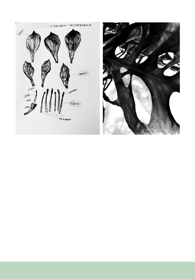
217
biological sciences (Baldwin and Crawford, 2012;
Flannery, 2012; Frei et al., 2010) help students
hone observational skills by creating works of art
that help them better understand connections
among, for example, the plant structure/function
concepts they are learning about, the details of
those structures they must “see” to draw, and how
those structures relate to functions. Exploring the
use of art as a learning mode in science is highly
regarded in some areas of biology, where faculty
have long recognized that developing drawing
skills to depict what one sees under the microscope,
in a dissecting pan, and in nature, allows students
to deepen their ability to see similarities and
differences in what they are studying (Klugman et
al., 2011).
Our course also brings to the fore the concept that
both artists and scientists are creatively engaged
in their disciplines and use similar modes and
practices. As noted by Beveridge in Seeds of
Discovery (1980): “The basic process of innovative
thinking is the same in science, the arts, business,
or any occupation that is not purely routine or just
following instructions.”
We meet for 5 hours each week: one 2.5-hour
class is in a biology lab and the other 2.5-hour
class is in an art studio. Lab time is spent learning
the structure and function of members of the
plant kingdom through lecture, lab, and field
observation. In the studio, students learn to
address a blank page creatively, and how to use
various media, such as graphite, pen and ink,
Figure 1. St. Catherine University Art major Blue Edwards created these two images for the Art of Biology class. The
image on the left is a lab sketch (pen and ink) of one of the flowers dissected in lab that day. On the right is Blue’s digital
drawing for the final creative art project—depicting a look through the holes in a Monstera leaf. Permission granted to
use images and student’s name and major.

218
and watercolor. We make explicit the connection
between the biological observation students do in
the lab and the drawing practice they complete in
the studio. The text we use, Botany for the Artist:
An Inspirational Guide to Drawing Plants by
Simblett (2010), complements what students are
learning in the lab and studio. Simblett reinforces
the connection between history, art, and biology
as she combines botanically correct and beautiful
drawings and paintings with descriptions of
plant organs, habitats, and instruction on how to
produce clear botanical illustrations that are also
works of art.
To enhance student learning, we invite local
artists, such as Marilyn Garber, Founder of the
MN School for Botanical Art, to teach classes, and
visit museum special holdings, such as the Maria
Sibylla Merian print collection at the Minneapolis
Institute of Art. Walks in nature, from our campus
full of flowering plants to the Eloise Butler
Wildflower Garden in Minneapolis, help students
understand the wide variety of plant structures
and how they evolved to facilitate a plant’s ability
to thrive, and gives them a chance to enjoy the
natural world.
In addition to a weekly sketchbook and quizzes
that test student knowledge of plant structure and
function and art concepts and practices, students
produce two exhibit-ready projects: one at mid-
term and one at the end of the semester, where
they combine what they have learned to create
pieces of art. These are presented at “openings”
complete with an artist’s reception, which is great
fun. The course is enjoyable to teach, and students
bloom (pardon the pun) throughout the semester
as they become more proficient in both art and
science.
ACKNOWLEDGMENTS
I would like to thank my writing group (Jane
Carroll, Joanne Cavallaro, Cecilia Konchar-Farr,
and Cindy Norton) for many years of thoughtful
and critical reading, great feedback, and for
friendship, support and encouragement.
REFERENCES
Baldwin, L., and I. Crawford. 2012. Does the science of
botany need art? Does art need the science of botany? CBA/
ABC Bulletin 45: 10.
Beveridge, W. I. B. 1980. Seeds of Discovery: A Sequel to
the Art of Scientific Investigation. Norton: New York, 130
pp.
Flannery, M. C. 2012. Biology & art: An intricate relation-
ship. American Biology Teacher 74: 194.
Frei, J., S. E. Alvarez, and M. B. Alexander. 2010. Ways of
seeing: Using the visual arts in nursing education. Journal
of Nursing Education 49: 672.
Hanriksen D. 2014. Full STEAM ahead: Creativity in excel-
lent STEM teaching practices. STEAM Journal 1(2).
Jemison, M. 2002. TED Talk. Teach Arts and Sciences
Together. Website: https://www.ted.com/talks/mae_jemi-
son_teach_arts_and_sciences_together
Klugman, C. M., J. Peel, and D. Blackmann-Menendez.
2011. Art rounds: Teaching interprofessional students visual
thinking strategies at one school. Academic Medicine 86:
1266-1271.
Quigley, C. F., D. Herro, and F. M. Jamil. 2017. Developing
a conceptual model of STEAM teaching practices. School
Science and Mathematics 117: 1–2.
Simblett, S. 2010. Botany for the Artist: An Inspirational
Guide to Drawing Plants. DK Publishing: New York. 256 pp
.
Snow C. P. 1961. The Rede Lecture, 1959: The two cultures.
Cambridge: Cambridge University Press.
Tott, C. D., T. L. Even, and S. M. Frame. 2020. Merging the
arts and sciences for collaborative sustainability action: A
methodological framework. Sustainability Science 15: 1067.

219
From the P SB Special I ssue on Art in the Botanical Sciences
It is the height of summer and you’ve ventured
out on your favorite trail. Late afternoon sun sets
the overhead leaves aglow, and the scent from
yesterday’s rain emanates from the soil beneath
your feet. A bird, plumed with vibrant stripes of
yellow and black, perches amid the pine boughs
and calls a looping, muddled chirp. You pause
in wonder, taking in this new sight and sound
among the otherwise familiar landscape. Taking a
notebook and pencil from your pack, you roughly
sketch the bird’s shape and a map of the woods
around you, jotting a few words to describe its
distinctive call. Another bird calls back, and you
note the number of birds joining the chorus. This
bridging of scientific observation and artistic
practice is nature journaling.
Nature journaling invites us to connect with
our curiosity for the natural world by keeping
records of our observations (Figure 1).
Nature Journaling: Sharing Perspectives
Between Art and Science
Corinn Rutkoski
1
Taylor Scamehorn
2
Robin Waterman
1
Misty Klotz
1
Meredith A. Zettlemoyer
1,3-4
1
W. K. Kellogg Biological Station, Hickory Corners, MI,
49060-9505
2
Kalamazoo Institute of Arts, Kalamazoo, MI 49007-
5102
3
Current address: Division of Biological Sciences, Univer-
sity of Montana, Missoula, MT 59812-0003
4
Corresponding author: meredith.zettlemoyer@umon-
tana.edu
Documentation can include words, numbers,
sketches, diagrams, photos, collage, maps, and
found objects. Anyone—even those not trained
in art or science—can use nature journaling to
cultivate mindfulness and wonder, build critical
thinking skills, develop greater perception, and
deepen their understanding of the environment.
Nature journaling instruction can be adapted
to any audience, from children curious about
basic processes in nature, to biology students
supplementing their academic training with real-
world observation, to those seeking to spend
quiet, intentional time in nature.
To share the practice of nature journaling, we
invite scientists, artists, and the public to engage in
an annual Nature Journaling webinar series. The
program was developed in 2020 as a partnership
between W. K. Kellogg Biological Station (KBS)
and the Kalamazoo Institute of Arts (KIA) with the
goals of building participants’ understanding of
ecology and evolution, developing nature drawing
skills, and encouraging journaling practices.
During each of four webinars, a KBS scientist
teaches an introductory ecology seminar focused
on a particular biological scale—landscapes,
communities, populations, or organisms—and
presents an example from their research. For
example, a lesson on landscape ecology includes
a case study on local habitat restoration (e.g.,
prescribed burning to maintain a native grassland)
whose goal is to connect patches of habitat for
native insect pollinators. A KIA artist then follows
with a brief lecture on art concepts that correlate
to the week’s themes and, using a photograph
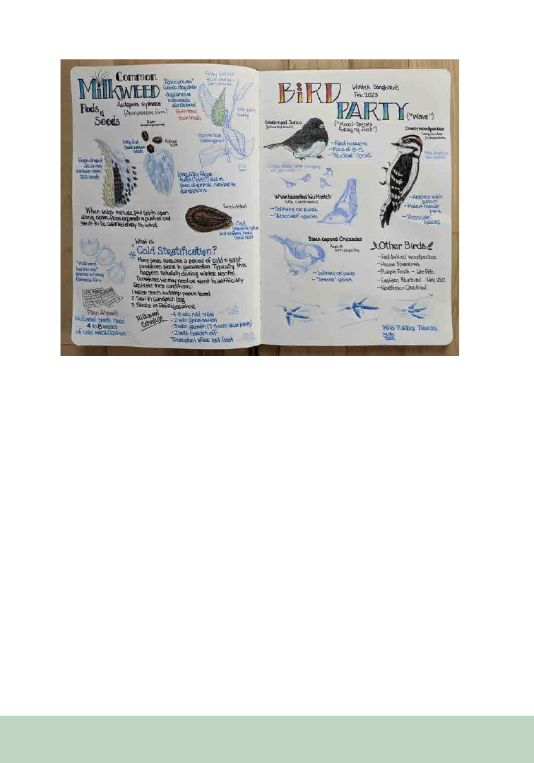
220
from the scientist’s research, guides participants
through an observational sketch of a landscape,
species interaction, or individual organism, each
week demonstrating a new technique. Participants
reflect on the activity and talk through challenges
they encountered.
Since 2020, we have partnered with ten scientists
and two artists who have presented a range of
ecology concepts and creative techniques in an
accessible format. Ecology presentations have
included topics such as prairie ecology and
management, the role of soil microbes in bioenergy
cropping systems, and moth species identification.
Art segments have incorporated art history, math,
and visual sciences to align instruction with each
week’s science theme. For example, following
a presentation on the mutualism between ants
and extrafloral nectaries, an artist demonstrated
that accurate proportions can be drawn by
interfacing multiple visualization techniques.
First, students were taught to reproduce correct
shapes from observation using their reference
image to measure lengths and a mutual angle at
two scales. Then, by learning to visualize negative
space, students effectively used geometric proofs
to test their proportions. The blending of these
two techniques demonstrated that in art, as in
science, the more data that can be synthesized, the
more accurate the results. Under the umbrella of
“mutualism,” this lesson pairing facilitated deeper
observation and understanding of the relationship
between things that rely on each other, whether
they are plants and insects, or lengths and angles.
Our four annual programs have reached 248
participants from eight states. The majority
of surveyed participants report improved
understanding of scientific concepts (81%) and
increased interest in starting or continuing their
nature journaling (95%). Participants deepen their
relationship with nature and art, are compelled to
be observant, and gain new knowledge about local
ecology in their own backyards.
Figure 1. Sample nature journaling page provided by 2023 participant Briar Hallowstone.

221
From the P SB Special I ssue on Art in the Botanical Sciences
KEYWORDS
animation, augmented reality, Caprifoliaceae,
interactive media, fusion, graphic design,
honeysuckle, Lonicera, science communication,
undergraduate education
Plants as a Case for Creative Collaboration: Designing the
Interactive Art-Science Exhibition
Meaningful Beauty
Christopher Ault
1,5
Wendy L. Clement
2,5
Anna Bergen
2
Michael J. Donoghue
3
Dianella G. Howarth
4
Leann Janzekovich
2
Siya Kakumanu
2
Robert King
2
Ria Shah
2
Mansa Srivastav
3
Shania Welch
2
Haley Wright
1
Jingbo Zhang
4
1
Department of Design and Creative Technology, The Col-
lege of New Jersey, Ewing, NJ 08628
2
Department of Biology, The College of New Jersey, Ewing,
NJ 08628
3
Department of Ecology and Evolutionary Biology, Yale
University, New Haven, CT 06520
4
Department of Biological Sciences, St. John’s University,
Jamaica, NY 11439
5
Co-authorship and corresponding author: ault@tcnj.edu,
clementw@tcnj.edu
Exploring Plant Diversity Through
Exhibit Design
From the perspective of a botanist, one might
describe honeysuckles (Lonicera, Caprifoliaceae)
as a group of nearly 140 species largely distributed
in the northern hemisphere, boasting an array of
diverse leaf, flower, and fruit forms. To the general
public in North America, however, awareness of
honeysuckle is usually limited to the sweet smells
of Lonicera japonica on summer walks through
the woods, or perhaps the tenacious creep of L.
morrowii into their yards and gardens. Inspired by
the ongoing research of a multi-institutional team
studying honeysuckle evolution—substantially
informed by the work of a graduate student at Yale
University and led by faculty at The College of New
Jersey (TCNJ)—a team of nearly 50 undergraduate
students collaborated to develop an art-science
exhibition that invited participants to view plants
through a variety of lenses. This collaborative
opportunity provided science communication
training opportunities for undergraduate students
in science, interactive media, art, and design.
Our students used their strengths to broaden
awareness of plant diversity and share novel
research outcomes on evolutionary studies of
honeysuckles through the design and display of
the exhibition. Ultimately named Meaningful
Scan this code with your
phone camera to reveal
augmented content through-
out the article.
Keeping your phone’s
browser open, scan
this and the other
black and white
symbols throughout
the story.

222
Beauty: The Vibrant Vocabulary of Honeysuckles,
the exhibition featured a rich blend of artistic and
scientific imagery, while employing digital and
interactive technology
to augment information
and personalize the experience for each
visitor.
The exhibition was first on display at the
TCNJ Art Gallery in Spring 2022, followed by the
Arnold Arboretum of Harvard University during
the sum
mer.
Project Development
The exhibition’s content,
design, and overall visitor
experience grew out of a
months-long collaboration
between student and faculty
scientists, artists, designers,
and creative technologists at TCNJ. Over the
course of the project, scientists and non-scientists
embraced the opportunity to share with each
other the knowledge, objectives, processes, and
tools of their respective disciplines. The primary
conduit for this collaborative work was a course in
Interactive Exhibit Design, offered by the college’s
Department of Interactive Multimedia, and taught
by Associate Professor Christopher Ault, as well
as a summer undergraduate research experience.
Interactive Exhibit Design assembled a diverse
group of student illustrators, animators,
programmers, graphic designers, writers, and
more. First offered remotely in the Spring of 2021
due to the pandemic, 16 students collaborated to
conceptualize various approaches to increasing
the public’s appreciation of honeysuckle diversity,
while also communicating specific insights
from the science team’s ongoing research on
honeysuckle evolution. Students received
institutional guidance from former TCNJ
students and the gallery directors from both
TCNJ and the Arnold Arboretum. The course
was offered again the following semester. Now
in person, the course’s 17 students had access
to the college’s “makerspace” to finalize designs
and fabricate the components of the exhibition.
A key improvement over the previous class
was to recruit two biology students as “learning
assistants.” As the interactive multimedia students
developed artwork, text, interactive interfaces,
and code, they could dependably turn to the
learning assistants to clear up any uncertainty
about the science. Some examples of this cross-
disciplinary peer teaching included deciphering
and creating floral diagrams, assisting navigation
of resources such as GBIF and iNaturalist, and
commenting on the visual interpretation of fusion
in honeysuckle leaves, flowers, and fruits. One of
the learning assistants—Biology student Robert
King—described the collaboration:
“Communicating with a group of students
who had little to no biology background was
a challenge, but it allowed me to increase
my science communication skills along with
my own understanding of honeysuckles. One
example was explaining to the artists the
orientation of flower morphology. Although I
was a scientist on the project—not an artist—I
am a visual learner, so I sketched all the flower
morphologies as part of the process. It helped
the artists understand the key concepts and
keep the botanical accuracy intact. It also
helped me appreciate the more than 140
species of honeysuckles.”
Between the two offerings of the course, the
exhibition made significant strides through a
summer research program at TCNJ that included
two interactive multimedia students and four
biology students. Anchored by weekly readings
and discussions of Beronda Montgomery’s Lessons
from Plants, both groups together considered some
of the factors behind plant visibility—or rather,
invisibility—on the part of the general public.
The science students on the team were challenged
to communicate their knowledge, methods,
vocabulary, and tools to the non-science students,
one of whom had a primary interest in animation,
and the other in user experience design. Armed
with these insights, the non-scientists conceived

223
of possible ways to communicate those ideas
through art, design, and technology. As the science
students came to understand both the goals of the
exhibition as well as the tools and processes of
their peers in interactive multimedia, they began
to share their own ideas for what approaches might
be effective for a gallery exhibition. This intensive
summer effort established the framework for the
entire exhibition and set goals for the exhibit
design course in t
he fall.
Interactive multimedia student Haley Wright
held the dual roles of project manager and major
contributing artist, while participating in both the
summer research program and the fall course. Of
the collaboration with science students, Wright
says:
“We gained a better understanding of the
bio team’s research by visiting their labs to
observe their work, examining honeysuckles
growing on campus, learning how to decipher
and create various diagrams, studying
images of live and pressed honeysuckle species
online, and discussing what we can learn
from honeysuckles and plants in general. We
pushed each other to think differently about
our work and how it can be communicated
to those outside of our field. As designers,
I believe this project helped prepare us for
working with a wide variety of clients in the
future, as it challenged us to infuse creativity
into a subject in which we were not an expert
ourselves and developed our ability to design
iteratively with the feedback of a client.”
Through the two semesters and summer, the
student collective generated a style guide—
including a color scheme, font, and tone—which
guided all decisions for development of all pieces
included in the gallery. Ultimately, the exhibit
was organized around six walls exploring themes
including regional diversity, morphological
diversity, and fusion through a combination of
illustration, animation, and graphic design made
interactive through augmented reality. Here we
describe how various forms of art were combined
with scientific content to sustain engagement of
visitors in an exhibition meant to communicate
the diversity of forms and function of plants.
Animations
Animation is a prominent area
of interest in the Interactive
Multimedia major, and several
students in the Interactive
Exhibit Design course
embraced the opportunity to
apply their skills to a project
that would be seen in person and at a large scale, as
opposed to a more typical platform like a YouTube
channel. Both animations were projected to fill
entire walls, using short-throw projectors to allow
visitors to view this “animated wallpaper” up
close without casting shadows (Figure 1D). After
considering various areas of the science that might
lend themselves to striking visuals, the students
arrived at two stylistically distinct approaches
that effectively blended science with an aesthetic
experience appropriate for the walls of an art
gallery.
One animation was inspired by herbarium
specimens, displaying black silhouettes of
fused honeysuckle leaves against bright colored
backgrounds. Elements of the silhouettes moved
and morphed, reminiscent of early abstract
animation where shapes were painted directly on
film stock. The other animation put into motion
original botanical illustrations from one of the
digital artists in the class, featuring honeysuckles
swaying in the breeze, with hummingbirds
and bees floating from flower to flower. Close
collaboration between the botanists, illustrators,
and animators led to a discussion on the nocturnal
nature of hawkmoth activity and how to portray
this in the wallpaper. As a result, when the scene
shifts from day to night, the hummingbirds
give way to hawkmoths, and fireflies blink like a
midsummer evening. At full size, the animation
was so striking that a group of visiting children
put their noses right up to the wall and wondered
aloud whether it was a movie or a painting.
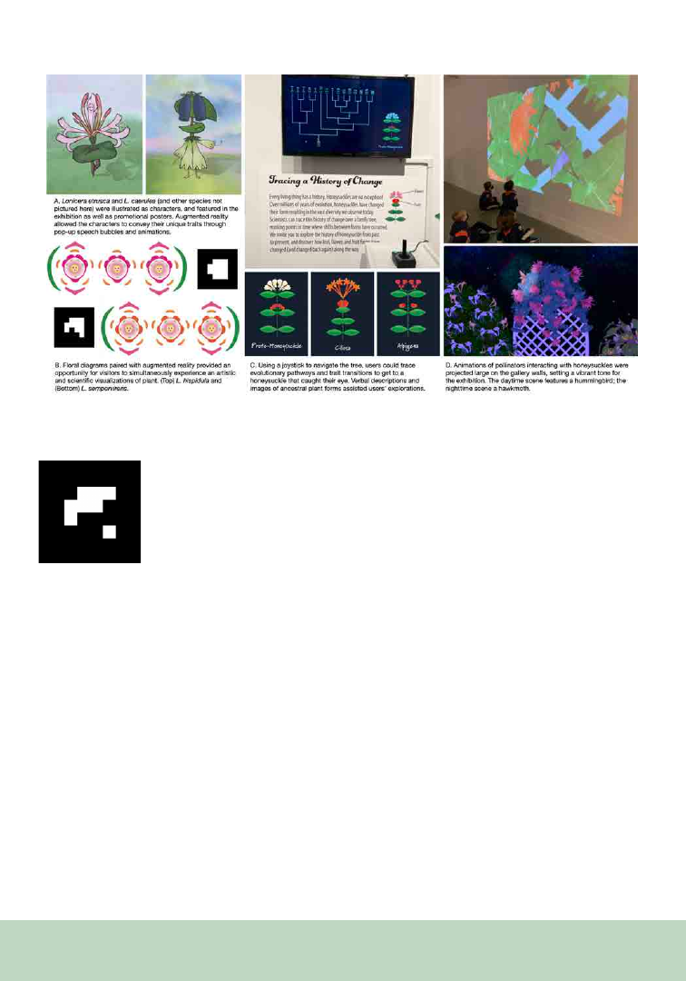
224
Augmented Reality
Augmented reality, commonly
shortened to AR, is a technique
by which digital, virtual content
is layered on top of a real-world,
physical environment. AR is
most commonly experienced
through phone screens—when a user scans a
special symbol with their phone’s camera, the
phone continues to display the “real” elements of
the scene visible to the camera, while integrating
digital content on top of it. This content might
range from 3D objects that appear to be actually
present in the space, to 2D text boxes offering
“pop up” information about one’s surroundings.
Early in the conceptual phase of the project, the
team recognized AR as a sort of magic lens—a
handheld translator—that would allow visitors to
move back and forth between the art and science
aspects of the exhibition. For example, when
a visitor scans a floral diagram of a particular
species, a 3D model of that plant extends out from
the wall in augmented reality (Figure 1B). When
a visitor scans a symbol on a playfully illustrated
tourist-style map of the United States, they’re
presented with a digital pop-up of essential facts
about honeysuckles common in that region of the
country. At the Arnold Arboretum, the team took
advantage of an opportunity not available at TCNJ,
placing AR symbols with corresponding species of
honeysuckle on the arboretum grounds, allowing
visitors to scan the symbols and learn more about
these plants.
Augmented reality was also key to one element of
the exhibition that aimed to establish a connective
throughline as well as provide users with hands-
on appreciation for the range of morphological
diversity in honeysuckles. Through a system
the team dubbed Build Your Own Honeysuckle,
visitors could scan AR symbols at key locations
throughout the exhibition and choose from a set
of options to create their own personalized, virtual
honeysuckle plants on their phones. Tied to the
theme of a particular wall—diversity, fusion,
plant communication, etc.—one AR location
would prompt visitors to choose the arrangement
of flowers on their plant, while another location
presented a choice of fused or unfused leaves. Once
Figure 1. Selected works from the Meaningful Beauty exhibition at The College of New Jersey
.

225
all choices were made, visitors scanned a final
symbol to see their unique virtual plant bloom
out of a real-world pot in a real-world garden
cart, surrounded by fanciful, abstract cut-outs
of honeysuckle-like shapes in other pots on the
cart. Designing the Build Your Own Honeysuckle
system demanded a balance of accurate science
communication with engaging user experience.
The interactive multimedia students worked back
and forth with the biology team to identify the key
elements that vary between species and endeavored
to portray these elements as accurately as possible
in a vibrant graphic style consistent with the
exhibition’s overall look and feel. After several
rounds of iterative design among the scientists
and graphic designers, the system included a total
of 144 different flower graphics.
Interactive Evolutionary Tree
Many students in the Interactive Multimedia major
are first and foremost interested in video game
design. And while they might not have enrolled
in the Interactive Exhibit Design course expecting
to apply those skills, the fact is museums and
galleries are meeting the demand and expectations
of their visitors by providing more interactive
content, often employing tools from game design.
In working to match students’ interests with
possible topics to explore, a phylogeny appeared
to lend itself to game design and would help
communicate novel outcomes from the research
of the graduate student, Mansa Srivastav. The
interactive evolutionary tree was the product
of close collaboration among undergraduate
scientists who studied these floral traits from living
plants, herbarium specimens, and the literature;
undergraduate graphic designers who created a
cartoon form of honeysuckles that exaggerated
the features that differentiated each species
without the loss of accuracy; and undergraduate
game designers who united these pieces with code
and a simple joystick inviting users as young as
4 years old to interact with the outcome (Figure
1C). In observing visitors, we found many sought
to find the path that led to a honeysuckle with a
certain set of features—perhaps pink flowers and
red fruits, or white flowers with small leaves, or
orange flowers and fused leaves. This suggests users
perhaps saw these plants as part of a pathway—
that is a tree marked by a series of changes along
an evolutionary trajectory culminating at one of
16 species. Our sense is that further development
of this particular design element could yield
additional innovations to provide users with a
much better grasp of phylogenetic trees and their
significance in interpreting evolutionary change.
Outcomes of the Exhibition
After two semesters of the Interactive Exhibit Design
course, the summer research program, and one
final flurry of work by the faculty, student project
manager Haley Wright and Gallery Director
Margaret Pazella-Granlund, the exhibition
opened at the TCNJ Art Gallery in February
2022. The exhibit was then reconfigured to fit
the multipurpose space in the Hunnewell Visitor
Center of the Arnold Arboretum the following
summer, under the guidance of Sheryl White,
Arnold’s Coordinator of Visitor Engagement and
Exhibitions.
Feedback from visitors clearly and consistently
suggested that the exhibition was effective in
terms of communicating the science, including
fostering understanding of honeysuckle diversity
and evolution, and that the artistic and interactive
elements of the exhibition served to increase their
understanding. Throughout the development
process, the team often discussed where the
exhibition fell on a spectrum from a natural
history experience to art experience, and visitors
expressed appreciation for our hybrid approach. A
tweet from one visitor offered:
Is it art?√
Is it science?√
Is it more than the sum of
art and science? √√√

226
As the students from both disciplines move on to
graduate school and/or positions in industry, they
do so having learned insights into the challenge
of conveying complex information in ways that
are appealing and engaging. Furthermore, they
can draw from an experience that demonstrated
first-hand the value of shared knowledge, diverse
perspectives, and earnest support between
collaborators. To our audience, the combination of
art and science gave permission to see and interact
with plants from a multitude of perspectives
that we hope increases their awareness and
appreciation of plant diversity worldwide.
ACKNOWLEDGMENTS
The authors would like to thank gallery directors
Margaret Pezalla-Granlund (TCNJ) and Sheryl
White (Arnold Arboretum). We also thank
Michael Dosmann and Kathryn Richardson for
access to and support for working in the living
collections at Arnold Arboretum, and Patrick
W. Sweeney for Lonicera photography. We thank
botanical collectors, arboretum, garden, and
herbarium curators as well as citizen scientists
for making the honeysuckles of the world more
accessible to all through their collections. Funding
was provided by TCNJ Center for the Arts, TCNJ
Cultural and Intellectual Community Council,
and National Science Foundation grants to W.L.C.
(DEB-1929670), M.J.D. (DEB-1929533), and
D.G.H. (DEB-1929674).

227
From the P SB Special I ssue on Art in the Botanical Sciences
KEYWORDS
anthropomorphism, belowground storage
organ, cuteness, functional traits, narrative,
pretense
Although plants are all around us, they are
complex, with many features difficult for humans
to perceive and understand. Thus, plant bodies
and ecology are challenging to explain quickly
and easily. Most focus on plants is aboveground,
but many perennial herbaceous plants need
belowground storage organs to recover from
seasonal dormancy or recurrent disturbance.
These organs are poorly understood by the
public and often
ignored or under-represented
by
scientists (Klimešová et al., 2020), perhaps
Understanding Plants Through Imagery:
Functional Traits, Cuteness, and Narrative
F. Curtis Lubbe
Institute of Botany of the Czech Academy of
Sciences, Dukelská 135, 379 01, Třeboň, Czech
Republic Email: curtis.lubbe@ibot.cas.cz
because they are hidden in soil. Belowground
storage organs store resources and coordinate the
growth and connection of aboveground stems and
leaves with belowground roots; in many ways they
are the core body of these plants. To really see and
understand the lives of plants, we must consider
the whole body of the plant and interpret the role
of their traits in plant growth and stress response
(Klimešová et al., 2020; Bartušková et al., 2022).
To explain plant life more clearly, we can give
plant belowground organs features and signs more
readily understood by humans. I will introduce
visual signs of the plant belowground and discuss
how anthropomorphized cuteness and narrative
can bring additional levels of meaning and
enjoyment. These tools can be used by anyone
trying to introduce or explain these topics to
colleagues or the general public, particularly in
more informal settings such as presentations,
posters, and blogs.
The Visible Plant
Belowground storage organs have great variety
in their size and traits, can be derived from roots,
stems, or leaves (all three main organ types),
and are frequently capable of clonal/vegetative
reproduction (Klimešová et al., 2017). Like
humans, plant bodies grow, move, and heal, but
they generally do this using belowground stems
that grow in units to branch and form different
structures that are lost and renewed annually
or over many years (Klimešová et al., 2017).
Belowground organs and their growth patterns
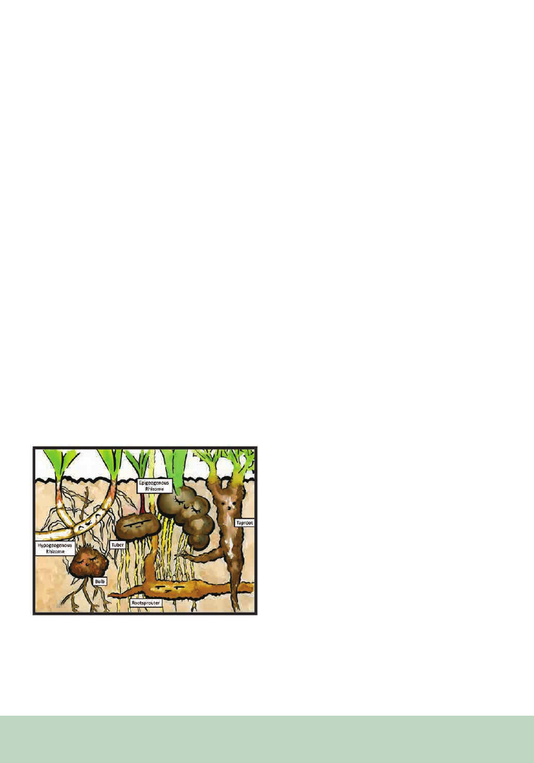
228
can be recognized by many different visible
characteristics (i.e., morphological functional
traits), although they are often obscured by soil,
roots, or their own complex structure (Bartušková
et al., 2022).
Plants generally move via growth, and the structure
of belowground plant bodies essentially depicts
behaviour and maintains the visual signs of life
history and response to the environment (Arber,
1950). Visible scars from previous stems can mark
age, and the shape of the body can show how it
has grown and moved in response to other plants
or obstacles (Raunkiær, 1934; Bartušková et al.,
2022). Plants can have round carbohydrate-rich
bodies for prolonged dormancy, long spreading
bodies that fragment and survive after damage,
or compact and tough bodies to survive drought
or cold (Klimešová et al., 2011, 2017; Bartušková
et al., 2022). To understand the complex lives of
plants, it is important to represent and even draw
attention to the belowground organs because
of their importance in plant life as well as their
hidden nature underground (Figure 1).
The Cute Plant
We can add cute features to images to make
plants and their lives more relatable and better
understood (Lubbe, 2022). Cuteness is visual
appeal in an endearing way and portrayed using
simplified imagery with the addition of signifiers
(see von Ehrenfels and Smith [1988] and Ashwin
[1984] for more on simplification and signifiers),
typically human qualities that can be used to
exploit human attitude toward anthropomorphism
(Lorimer, 2007). Anthropomorphism is the use of
human qualities inferred onto non-human subjects
(Epley et al., 2008), including body shape, limbs,
activities, and recognizable facial expressions
and emotions (Duffy, 2014). Anthropomorphic
interaction is an extension of relationship and
empathy to a non-human subject, often used to
cope and relate in the real world (Airenti, 2018).
This can be held by people as a way of relating to
outside subjects (Severson and Woodard, 2018)
and can bring comfort and understanding (Epley
et al., 2008; Airenti, 2018).
Cuteness is most often applied to material that is for
children or entertainment and may be best used in
more informal settings, but when carefully applied
(Geerdts et al., 2016), these signifiers can convey
useful information that could help any audience
understand a topic (Chan, 2012). For example, a
face on a plant storage organ can indicate its role as
a body, and a sad expression can indicate an effect
from damage or suboptimal conditions (Figure
2; Bruni et al., 2018). The compact and rounded
belowground bodies of many plants adapted for
stressful environments can make cuteness easier
to convey and their situations are more dramatic,
allowing for lively expressions. Designers
frequently take advantage of the shapes and body
arrangements very different from humans to make
creative and appealing images (Duffy, 2014).
Figure 1. Diversity of belowground storage organs. Rhizomes
and tubers are stem derived. Bulbs are short stems with leaves
for storage. Taproots are stem-tissue with a large root below.
Rootsprouters are roots adapted to form new stems.
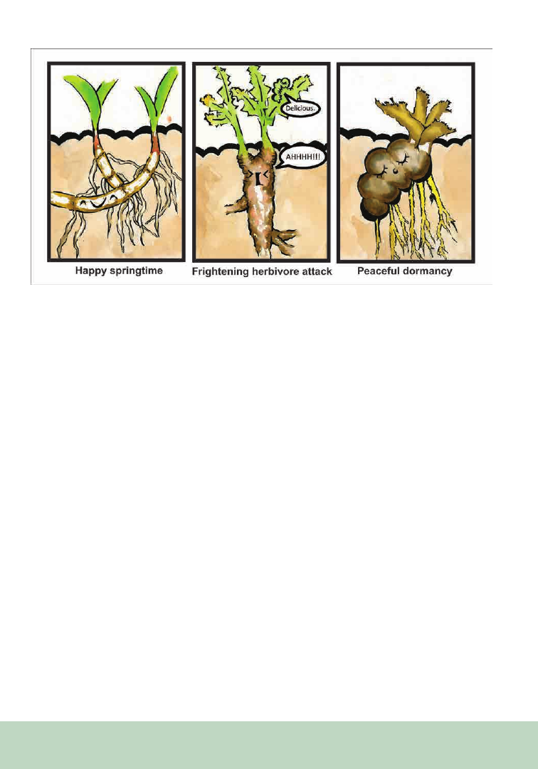
229
The Cute Plant Tells Its Story
Narrative is another powerful tool to communicate
information and increase understanding, which
is especially important to explain concepts more
distant from the human experience (Negrete and
Lartigue, 2004; Dahlstrom, 2014; Lin et al., 2015).
Anthropomorphism may aid in forming the
subject as a relatable and recognizable character
for use within narrative (Farinella, 2018) and bring
levity and lightheartedness to a subject to engage
the viewer (Yeo et al., 2020). Anthropomorphic
features and imagery are also part of pretense/
pretending, holding counterfactuals as imaginative
play or amusement, commonly used by children
to learn and grow (Severson and Woodard, 2018).
Anthropomorphism and pretense can increase
enjoyment, engagement, understanding, and
retention of details for both children and adults
(Geerdts et al., 2016; Yeo et al., 2020; McGellin et
al., 2021). These tools can also be used to correct
possible misconceptions and illuminate obscure
topics (Chan et al., 2012), which may be especially
useful for understanding perennial herbs because
they have a diverse range of belowground
storage organs that form their central bodies,
all hidden in the soil. By making narratives that
include the belowground organs of plants (and
their informative traits) and including our own
expressive features, we can thus see and relate to
the full lives of plants (Figure 3).
Conclusions
The visual signs of plant functional traits, human
expressions, and narratives can all convey
information about plant life and be fun at the
same time. To learn about the lives of plants, we
must dig them up, see these signs both above and
below the soil, and show them to people to give
greater learning and understanding. Cuteness
and narrative are not mandatory, but they can
increase understanding and give viewers a more
personalized view of how we view plants and the
enjoyment we have in our work.
Figure 2. Plant expressions.
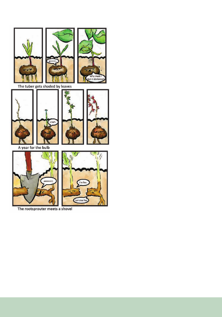
230
ACKNOWLEDGMENTS
This manuscript was supported by the long-
term research development project of the Czech
Academy of Sciences [No. RVO 67985939] and by
the Praemium Academiae award from the Czech
Academy of Sciences of the Czech Republic.
REFERENCES
Airenti, G. 2018. The development of anthropomorphism
in interaction: intersubjectivity, imagination, and theory of
mind. Frontiers in Psychology 9: 2136.
Arber, A. 1950. The natural philosophy of plant form. Cam-
bridge University Press.
Ashwin, C. 1984. Drawing, design and semiotics. Design Is-
sues 1: 42.
Bartušková, A., F. C. Lubbe, J. Qian, T. Herben, and J.
Klimešová. 2022. The effect of moisture, nutrients and dis-
turbance on storage organ size and persistence in temperate
herbs. Functional Ecology 36: 314–325.
Bruni, D., P. Perconti, P., and A. Plebe. 2018. Anti-anthropo-
morphism and its limits. Frontiers in Psychology 9: 2205.
Chan, A. A. Y.-H. 2012. Anthropomorphism as a conserva-
tion tool. Biodiversity and Conservation 21: 1889–1892.
Dahlstrom, M. F. 2014. Using narratives and storytelling to
communicate science with nonexpert audiences. Proceed-
ings of the National Academy of Sciences 111 (supplement_4):
13614–13620.
Duffy, B. 2014. Anthropogenic anthropopathous anthropo-
morphic USPTO trademarks: The Plant People phenomenon
model of anthropomorphism. In S. Brown, S. Ponsonby-
McCabe (eds.), Brand mascots and other marketing animals,
219–219. Routeledge.
Epley, N., A. Waytz, S. Akalis, and J. T. Cacioppo. 2008. When
we need a human: Motivational determinants of anthropo-
morphism. Social Cognition 26: 143–155.
Farinella, M. 2018. The potential of comics in science com-
munication. Journal of Science Communication 17: Y01.
Geerdts, M. S., G. A. Van de Walle, and V. LoBue. 2016.
Learning about real animals from anthropomorphic media.
Imagination, Cognition and Personality 36: 5–26.
Klimešová, J., J. Danihelka, J. Chrtek, F. de Bello, and T. Her-
ben. 2017. CLOPLA: a database of clonal and bud-bank traits
of the Central European flora. Ecology 98: 1179.
Figure 3. Plant stories.

231
Klimešová, J., J. Doležal, and M. Sammul. 2011. Evolution-
ary and organismic constraints on the relationship between
spacer length and environmental conditions in clonal plants.
Oikos 120: 1110–1120.
Klimešová, J., J. Martínková, G. Ottaviani, and T. Charles-
Dominique. 2020. Half of the (big) picture is missing! Ameri-
can Journal of Botany 107: 385–389.
Lin, S. F., H. S. Lin, L. Lee, and L. D. Yore. 2015. Are science
comics a good medium for science communication? The case
for public learning of nanotechnology. International Journal
of Science Education, Part B 5: 276-294.
Lorimer, J. 2007. Nonhuman charisma. Environment and
Planning D: Society and Space 25: 911–932.
Lubbe, F. C. 2022. A Call for Cuteness: Imagery in Science
Communication and Outreach. In Nature Portfolio Ecology
& Evolution Community. Website: https://ecoevocommunity.
nature.com/posts/a-call-for-cuteness-imagery-in-science-
communication-and-outreach.
McGellin, R. T. L., A. Grand, and M. Sullivan. 2021. Stop
avoiding the inevitable: The effects of anthropomorphism in
science writing for non-experts. Public Understanding of Sci-
ence 30: 621–640.
Negrete, A., and C. Lartigue. 2004. Learning from education
to communicate science as a good story. Endeavour 28: 120-
124.
Raunkiær, C. 1934. The life forms of plants and statistical plant
geography. Oxford University Press.
Severson, R. L., and S. R. Woodard. 2018. Imagining others’
minds: The positive relation between children’s role play and
anthropomorphism. Frontiers in Psychology 9: 2140.
von Ehrenfels, C., and B. Smith. 1988. On Gestalt qualities.
In B. Smith (ed.), Foundations of Gestalt Theory (pp. 11–8).
Philosophia.
Yeo, S. K., L. Y.-F. Su, M. A. Cacciat , M. McKasy, and S. Qian.
2020. Predicting intentions to engage with scientific messages
on twitter: The roles of mirth and need for humor. Science
Communican 42: 48107.
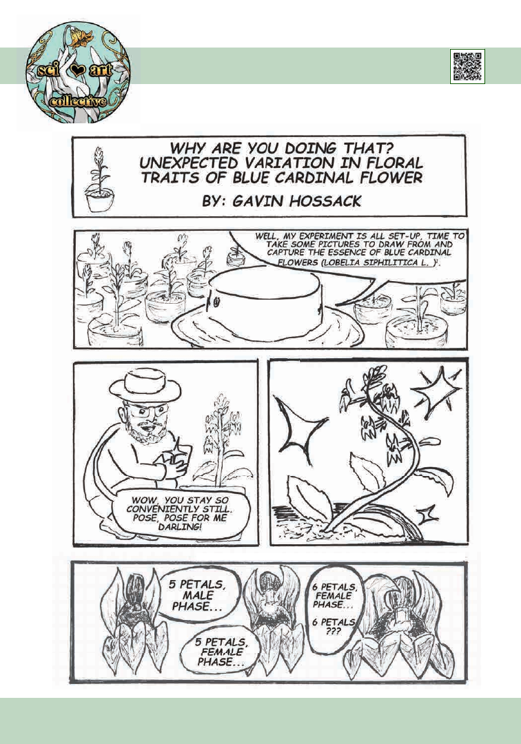
232
From the P SB Special I ssue on Art in the Botanical Sciences
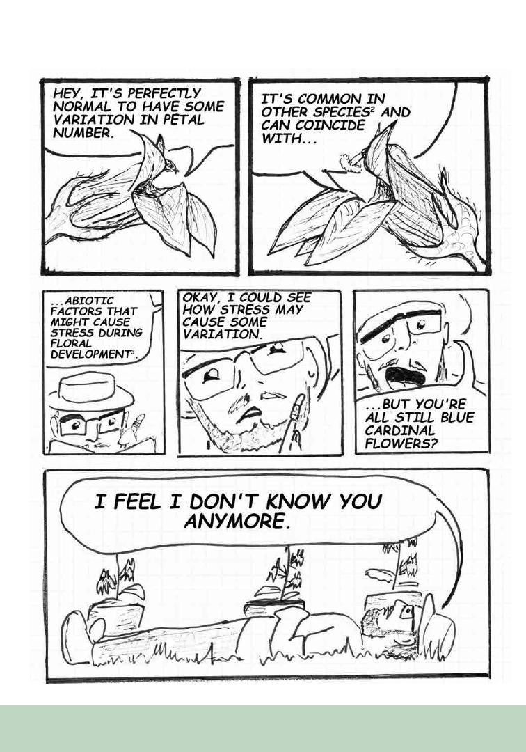
233
139
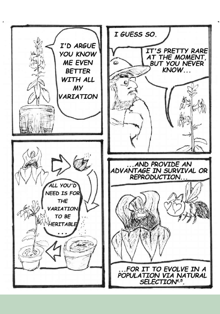
234
140
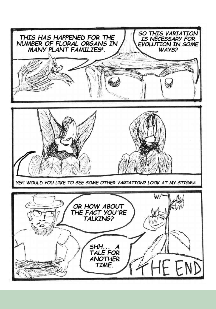
235
141

236
REFERENCES
1
Hossack, G. C. and C. M. Caruso. 2023. Simulated
pollinator decline has similar effects on seed produc-
tion of female and hermaphrodite Lobelia siphilitica,
but different effects on selection on floral traits. Ameri-
can Journal of Botany 110: e16106.
2
Ellstrans, N. C., and R. J. Michell. 1988. Spatial and
temporal patterns of floral inconsistency in plants and
populations of Ipomopsis aggregata (Polemoniaceae).
Botanical Gazette 149: 209–212.
3
Huether, C. A. 1969. Constancy of the Pentamerous
Corolla phenotype in natural populations of Linanthus.
Evolution 23: 572–588.
4
Conner, J. K., A. M. Rice, C. Stewart, and M. T. Mor-
gan. 2003. Patterns and mechanisms of selection on a
family-diagnostic trait: evidence from experimental
manipulation and life-time fitness selection gradients.
Evolution 57: 480–486.
5
Mickley, J. G. 2017. The adaptive nature of stasis
for petal number: can pollinator-mediated stabilizing
selection explain five-petaled flowers? Ph.D. disserta-
tion, University of Connecticut, Storrs, CT, USA.
6
Roonse De Craene, L. 2016. Meristic changes in
flowering plants: How flowers play with numbers.
Flora 221: 22–37.
Gavin Hossack
Port Perry, Ontario Canada
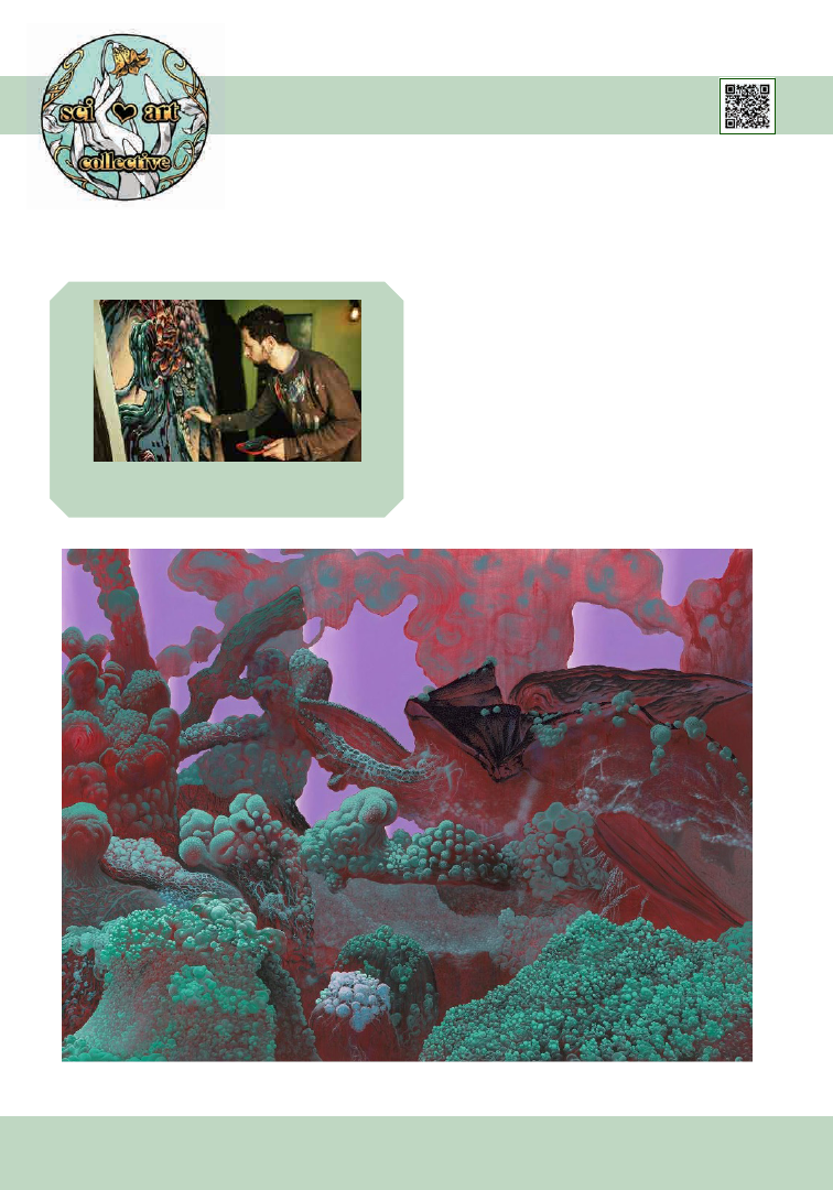
237
From the P SB Special I ssue on Art in the Botanical Sciences
I am a painter and a graduate of the Bezalel
Academy of Art and Design. I have my own studio
in Rechovot, Israel, where I paint my personal
art. I also do wall paintings, and live painting
performances.
The painting shown in Figure 1 is part of a series
called substrate, in which I paint visions from a
small bowl containing soil fungi and slime mold
that I grew. The bowl serves as the central stage
for the unplanned wild growths from my pots at
home; in this sense, it is a tiny uncivilized jungle
Wild Growth Paintings
Daniel Philosoph
Figure 1. Daniel Philosoph, Substrate 6, oil and acrylic on canvas, 250×180 cm, 2020.
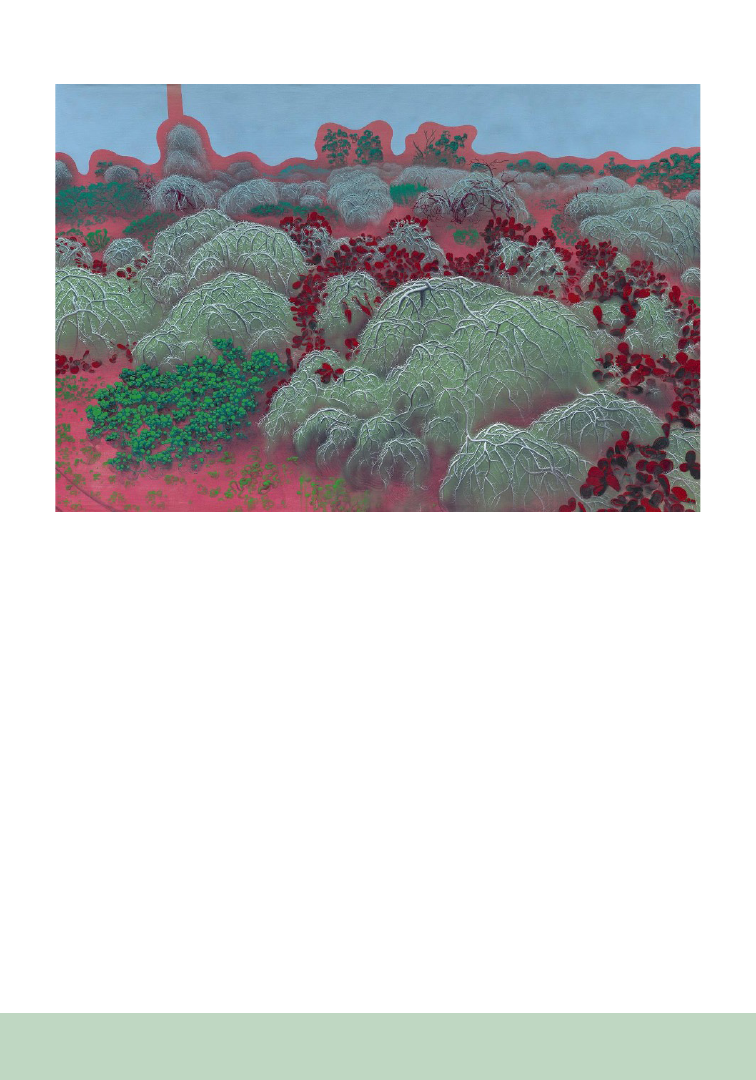
238
growing in my civilized bowl. In this painting I
looked through a window of about 2 × 1.5 cm from
a side angle that creates a view of a landscape. The
observation is done through different macroscopic
magnifying glasses, which produces multiple
perspectives and focuses on every detail in the
painting—a combination of depth and flattening.
The painting shown in Figure 2 is a “far-out.” I
mean that I treat it like a close-up painting, only in
reverse. This is a piece of landscape located at the
edge of the city where wild crops have taken over
what was once a field/cultivated area and is now
abandoned, allowing the wild growth of invasive
vegetation. It is a type of an unplanned jungle
that reclaims its grip on the ground using leftover
remnants. This painting is based on a photo I took
with a drone, and it simulates a magnifying-glass view.
In these paintings I look at the wild biology that
is at the edge of my existence, the edge of my
flowerpot, or my city. These are multi-layered
process paintings that try to draw from the visual
of the wild biology and create a parallel process
of the growth of the painting on the canvas. This
is done by breaking down the visual into different
actions that represent parts in visibility and
structure (the light, the branches, the parasitic
plant, the biology that serves as a growing
medium, etc.). Each operation uses a certain
shade or two, a certain dilution, and a different
character. Any such operation is blind, and as
soon as its properties are determined, it runs over
the painting and rises above the previous layers.
The painting is the result of all the processes that
took place on the canvas—a kind of biology of a
painting.
In my work, I draw inspiration from the way in
which science examines biology and describes
the processes that take place. This biological
information serves me, as a kind of internal,
subconscious information that exists in nature
and provides me with another perspective on the
sights of nature.
Figure 2. Daniel Philosoph, On the way to Mayan, oil, acrylic, and spray on canvas, 180×120 cm, 2020.

239
From the P SB Special I ssue on Art in the Botanical Sciences
Sometimes a book changes the trajectory of your
life. Frederick Case’s Orchids of the Western Great
Lakes Region was that turning point for me. There
are 40+ species of orchids in the Chicago region??
What a revelation! But how were they doing now?
My son was collecting Boy Scout merit badges at
the time, and we used that as an excuse to visit
natural areas and look for orchids and to learn
more about where they lived. The naturalist
leading a tour at Volo Bog told us how she had
found a Rose Pogonia (Pogonia ophioglossoides)
lying on the boardwalk, plucked up and then cast
aside. “How could they?!” I thought. A lack of
deep awareness and empathy for nature seemed
to be commonplace, so was it possible to make a
difference somehow? Synchronistic events led to
enrollment in Principles and Practices of Rare
Plant Monitoring, with Susanne Masi, one of the
founders of the North Branch Restoration Project
in Cook County, IL, and then co-founder of the
Chicago Botanic Gardens’ Plants of Concern
program. That led to becoming part of the natural
Art + Botany: Making a Difference
Kathleen Marie Garness
Scientific affiliate, Field Museum
Science and conservation affiliate,
Morton Arboretum
area’s stewardship and restoration community,
then to painting midwestern orchids and teaching
others about them.
One dilemma most stewardship volunteers have
is identifying the plants in their preserves. How
could you tell the invasive species you needed to
remove, from the native ones who had evolved
there? Field botany is seldom taught in colleges
anymore, much less high schools, and popular
field guides are incomplete. Most of us had
little familiarity with scientific (dichotomous)
keys—those were considered the province of
the elite botanist. During restoration workdays,
experienced stewards would point out a plant, tell
us its common and scientific names, with the hope
that we would remember them at next encounter.
There seemed to be no middle ground between
generic botanical guides and exhaustive keys such
as those found in Gray’s Manual of Botany (Fernald,
1950) or Gleason and Cronquist’s
Manual of
Vascular Plants of Northeastern United States and
Adjacent Canada. How can one appreciate and
protect biodiversity if one cannot recognize it?
How do you shift from seeing masses of vegetation
into seeing this dazzling array of plants as friends
and companions on your journey who you’d like
to get to know better?
Although the iNaturalist app has become
increasingly widespread and may help with some
identifications, there are many situations where
you simply have to use a dichotomous key and
invest the time and work in order to learn how to
do it. Admittedly, that is an advanced step. Can
there be a middle ground that is scalable?

240
Twelve years ago, Cook County steward Barbara
Birmingham asked me to help her develop some
illustrated plant family pages for her volunteers.
Collaborations with professional botanists and
ecologists were necessary so the pages could be
both accurate and succinct. Other pages followed.
The Field Museum offered to publish them,
seeing they met a wider need to learn about
our diverse flora here in the Chicago region.
The free download, Common Plant Families of
the Chicago Region, introduces students to 23
regional plant families, including examples of
non-native invasive species that threaten remnant
ecosystems. Other guides followed, including an
illustrated glossary to the regionally respected
Flora of the Chicago Region as well as several other
genus-level treatments published by Conservation
Research Institute. There are so many features
covered in dichotomous keys, it is important to
have a visual reference for when you run into a
new term. And there is really no substitute for that
level of study, nor should there be. Working with
Flora of the Chicago Region co-author Dr. Gerould
Wilhelm and other botanists taught me that to
draw plants accurately, you first must take time
to observe them very carefully, then read the keys
carefully, drawing only what is essential to see how
that species differs from those similar. I worked
primarily from herbarium material.
With over 7000 downloads by 2019, Common
Plant Families of the Chicago Region was showing
its usefulness. The joint Chicago Botanic Garden/
Northwestern University masters and doctoral
programs, the University of Illinois Master
Naturalists’ program, the Forest Preserves of Cook
County, and local restoration contractors continue
to use it to train students, staff, and interns.
As of June 2023, the guide has been translated
and downloaded 13,826 times in the English
version, 4831 times in Spanish, and 172 times in
Portuguese.
Speaking
recently with a colleague tasked with
hiring ecologists for a regional forest preserve
program, she recounted how many applicants
with Masters degrees in ecology knew only a
few native plants and their plant community
associates, and appeared flummoxed when asked
what they knew about plant taxonomy. This lack
of plant knowledge is disqualifying them for good
jobs in the profession. The situation is even worse
in the growing field of natural areas restoration
contractors, who need to know the difference
between the native Purple Prairie Clover (Dalea
purpurea) and purple Blazing Stars (Liatris spp.)
from the non-native, invasive Purple Loosestrife
(Lythrum salicaria), and who are set loose with
chemicals and only rudimentary training in plant
identification. This ought to be avoidable, but I
hear this story again and again. It is our hope these
guides and others will make a difference.
Another question surfaced: how do we reach out
beyond our rather insular, aging, community
of well- educated people, mostly of European-
American descent, involved with botany and
natural areas restoration work? How about
through art? Art is a universal language. In 2013
the American Society of Botanical Artists funded
our project to bring botanical illustration to new
and diverse audiences, supplying materials to over
70 students of all ages, ethnicities, and abilities,
in ten regional venues: nature centers, forest
preserves, and nature museums. In 2022 they
funded our research based around Illinois Beach
State Park, to document and illustrate the 23
species of orchids identified there from herbarium
records. The Volunteer Stewardship Network of
the Illinois Nature Conservancy provided funding
for more art materials for three children’s classes,
one for teens, and two for adults in Zion and
Waukegan, IL, where almost 50% of the residents’
primary language is Spanish.
Art is a way of seeing things more deeply and
clearly. In order to draw a plant accurately, you
need to study all its parts, observe it from all angles,
touch it, smell it, and develop a relationship with
it. Old botany textbooks featured drawing plant
parts at each level of the curriculum. We hope
to recapture some of the wonder and curiosity of
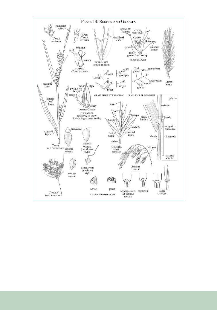
241
childhood in each of these lessons, to help others
experience the beauty and complexity of the plant
world and find inspiration in it.
There
is a spiritual thread in all of this—a quest
to find meaning and beauty in the world, to
deeply engage with our native flora. We know that
nature is a refuge for our spirits; during COVID,
people flocked in droves to the forest preserves
and other natural areas, in visitation numbers not
seen in many years. But do the visitors make the
connection between how they feel out in nature
with nature’s pressing need for compassionate
stewardship? The goal of my art, therefore, and
especially the teaching of botanical art, is to engage
people with plants—to see them more precisely, to
learn the language of botany, and to identify and
describe what they see. And learn to care for them
and participate in millennia-long human-cultural
relationships that will benefit all of us.
REFERENCES
Fernald, M. L. 1950. Gray’s Manual of Botany, ed 8.
Dioscorides Press: Portland, OR. 1632 pp.
Figure 1. Sedges and grasses.
PSB 69 (3) 2023
242
��������������������<�/p>
Amesbury, MA 01913 U.S.A. | +1 978-834-0505 | sales@ppsystems.com
Learn more about the CIRAS-4
Portable Photosynthesis System
The fastest, most accurate leaf gas exchange system
available that is elevating the high-level field
research experience worldwide.
Schedule a virtual demonstration today!
High-level field research
demands fast-response
technology, precise data,
and true portability.
• Designed for high-level field research
Lightweight, compact & up to 16 hours of
continuous use
• Fully automatic & programmable
environmental control
Full control of CO
2
, H
2
O, temperature &
light (RGBW-FR)
• Exceptionally intuitive, customizable
software
So easy to learn & use. Begin taking
measurements right out of the box
• Measure rapid A/C
i
in real time without
post-processing
The Single-Step CO
2
Response (SSCO
2
R
™
)
Method is the fastest, most accurate,
streamlined method available
• Only system to feature far-red LEDs
Control far-red light up to 30% of PAR
• Measure leaf gas exchange & chlorophyll
fluorescence simultaneously
Plus dark & light-adapted fluorescence
measurement parameters & OJIP fast
induction kinetics
PSB 69 (3) 2023
243
OUP Plant Science Hub
Bringing together hundreds of articles from across all our
plant science journals, organized thematically for the first time.
Plant Resilience
Sustainable Plant Production
Developing Technologies
Plant Science to Improve Health
Explore the topic hubs now >>

PSB 69 (3) 2023
244
244
SOCIETY NEWS
Dr. Thomas Givnish is an internationally
renowned scientist who is acclaimed for
his “sharp intellect, breadth of knowledge,
creativity, and productivity.” He is known
for his unique interdisciplinary research
Botanical Society of America’s
Award Winners (Part 2)
Distinguished Fellows of the Botanical Society of America
The “Distinguished Fellow of the Botanical Society of America” is the highest honor our
Society bestows. Each year, the award committee solicits nominations, evaluates candidates,
and selects those to receive an award. Awardees are chosen based on their outstanding
contributions to the mission of our scientific Society. The committee identifies recipients who
have demonstrated excellence in basic research, education, public policy, or who have pro-
vided exceptional service to the professional botanical community, or who may have made
contributions to a combination of these categories.
Dr. Thomas Givnish
University of
Wisconsin-Madison
and for his breadth of expertise, making
major contributions to such diverse areas as
ecophysiology, systematics, biomechanics,
plant-animal interactions, adaptive radiation,
and species diversification and extinction.
Over the past 51 years, Tom has authored,
coauthored, or edited more than 160 papers,
books, and book chapters, which have been
cited more than 21,000 times. And he has
sustained substantial funding from the
National Science Foundation to support his
research for many years.
Tom has made foundational contributions in
the fields of ecology and systematics: “his work
on the evolution of monocots, bromeliads,
and carnivorous plants, among other groups,
has been equally as transformative as his
experimental and theoretical studies on plant
functional traits, plant height, leaf form, and
photosynthetic physiology.” Tom’s early work
in plant ecology was grounded in his keen
interest and background in mathematics
and economics. He developed mathematical
models inspired by economic theory to
PSB 69 (3) 2023
245
explore and explain the adaptive nature of leaf
form and plant height. He was an early adopter
of molecular techniques for studying ecology
and evolution, and he blended his work on
adaptation with phylogenetic resources,
applying the concept of adaptative radiation
to plants. His 1997 book Molecular Evolution
and Adaptive Radiation, co-edited with Ken
Sytsma, remains authoritative. His work in
phylogenetic systematics, and his embrace of
new methods and approaches, has kept him
at the forefront of developments in the field.
Tom’s focus on monocots has transformed our
understanding of the evolutionary history and
higher-level relationships of that group. And
he’s showing no signs of slowing down.
Tom has immense and infectious enthusiasm
for both fieldwork and the lab, and his
knowledge of plant diversity, natural history,
and ecology is encyclopedic and insightful.
He shares his knowledge and passion for
plants with his students and colleagues and
reaches beyond the University as well through
the “Wednesday Night @ the Lab” televised
outreach talks, presentations to naturalist and
gardening groups, and engagement with local
conservation groups. He is also involved in the
DNR Citizens Advisory Board for Wisconsin
Dells Natural Area and the U.S. Fish &
Wildlife Service National Recovery Team for
the Karner Blue butterfly. He is a botanist
well-deserving of the BSA Distinguished
Fellow Award.
Dr. Steven Manchester
Florida Museum of Natural History
Dr. Steven Manchester is one of the world’s
leading specialists in fossil plants, whose
research has had “a profound impact on the
directions in the scientific exploration of
angiosperm diversification and biogeographic
patterns, particularly through the Cenozoic.”
Through extensive field work throughout
the world, including in the western US,
eastern Asia, India, Europe, and Panama, and
through careful study, he has documented
some of the earliest known fruits for several
families of flowering plants, including the
banana (Musaceae), kiwi (Actinidiaceae),
grape (Vitaceae), and walnut (Juglandaceae)
families. Dr. Manchester often works at the
“interface of the living and the dead, working
closely with angiosperm systematists to
integrate fossils into phylogenetic trees of
living species.” He also has worked with
molecular systematists to provide fossil
calibration points for molecular dating
analyses. His broad network of international
collaborations has included researchers from
all career stages and backgrounds, and he is
known to be generous with his time and ideas.

PSB 69 (3) 2023
246
BSA CORRESPONDING MEMBERS AWARD
Corresponding members are distinguished senior scientists who have made outstanding
contributions to plant science and who live and work outside of the United States of America.
Corresponding members are nominated by the Council, which reviews recommendations and
credentials submitted by members, and elected by the membership at the annual BSA business
meeting. Corresponding members have all the privileges of life-time members.
Dr. Gonzalo Nieto Feliner, Royal Botanical Garden of Madrid
AWARDS FOR ESTABLISHED SCIENTISTS
GIVEN BY THE SECTIONS
CONTRIBUTIONS TO PALEOBOTANY AWARD
Paleobotanical Section
Ruth A. Stockey, Oregon State University
In addition to his impressive publication
record, his many invited presentations and
research grants from NSF and other funding
sources, Steve has also been deeply engaged in
public outreach engaging amateur collectors
and students in his field campaigns. He is
described by several colleagues to be hard-
working, with a passion for field work, which
he considers essential to paleobotanical
research. He is also described as kind,
thoughtful, modest, having a great sense of
humor, and intensely dedicated to student
training and mentorship.
Steve is a life member of both the Botanical
Society of America and the American
Society of Plant Taxonomists. He has held
leadership roles in the Paleobotanical Section
of BSA and the International Organisation
of Palaeobotany (IOP), in which he served
as President for many years. He is a Foreign
Representative Member of The Gondwana
Geological Society based in Nagpur, India,
and is active in the Association of Wood
Anatomists and the American Association
of Stratigraphic Palynologists. He has served
on his department’s Natural History Advisory
Committee and chairs his institution’s
IDEA (Inclusion, Diversity, Equity, and
Accessibility) Committee, which recently
developed an endowed scholarship fund for
graduate students of minoritized groups and
an internship program for students from
groups that are underrepresented in science in
the US. He is a great model for service to the
professional community and well deserving of
the BSA’s Distinguished Fellow Award.

PSB 69 (3) 2023
247
EDGAR T. WHERRY AWARD
Pteridological Section and the American Fern Society
The Edgar T. Wherry Award is given for the best paper presented during the contributed papers
session of the Pteridological Section. This award is in honor of Dr. Wherry’s many contributions to
the floristics and patterns of evolution in ferns.
Katelin Burow, Purdue University, For the Presentation: Genetic Mechanisms of Sex
Determination in Ceratopteris richardii. Co-authors: Grace Estep, Brian Dilkes, Jody Banks,
Jen Wisecaver.
Sonia Molino, Universidad Complutense de Madrid, For the Presentation: Discovering
Parablechnum: a complex evolutionary history within the youngest fern family. Co-
authors: Weston Testo, Mario Mairal, Guillermo Santos-Rivilla, Rafael Medina
MARGARET MENZEL AWARD
Genetics Section
T
he Margaret Menzel Award is presented by the Genetics Section for the outstanding paper presented
in the contributed papers sessions of the annual meetings.
Lauren Frankel, University of Wisconsin-Madison, For the Presentation: Summary tests of
introgression are highly sensitive to rate variation across lineages. Co-author: Cécile Ané
MICHAEL CICHAN PALEOBOTANICAL
RESEARCH GRANT
Paleobotanical Section
The Award is to provide funds for those who have completed a PhD and are currently in a
post-doctoral position or non-tenure track position.
Ana Andruchow-Colombo, University of Kansas, For the Paper: Placing the Voltziales: A
study of the origin and evolution of modern conifers.
Michael D’Antonio, Field Museum, For the Paper: Reconstruction and systematics of conflictive
late Paleozoic plants using tomography and microphotography on Mazon Creek nodules and
coal balls.

PSB 69 (3) 2023
248
AWARDS FOR STUDENTS
THE BOTANY AND BEYOND: PLANTS GRANTS RECIPIENTS
The PLANTS (Preparing Leaders and Nurturing Tomorrow’s Scientists: Increasing the diversity of
plant scientists) program recognizes outstanding undergraduates from diverse backgrounds and
provides travel grant.
Nico Andrade, University of Florida, Advisor: Drs. Pam and Doug Soltis
Sofia Baez, Old Dominion University, Advisor: Lisa Wallace
Cari DeCoursey, Weber State University, Advisor: Dr. James Cohen
Fitzwilliam Dettmer, Rutgers University New Brunswick, Advisor: Dr. Lena Struwe
Natalie Heaton, University of Florida, Advisor: Lucas Majure
Chinyang Huang, Purdue University, Advisor: Dr. Daniel Park
Anij Mackey, Texas Tech University, Advisor: Matthew G. Johnson
Marife Minaya, California State Polytechnic University, Pomona, Advisor: Carrie Kiel
Hashel Orquiz, University of Texas at El Paso, Advisor: Dr. Michael Moody
HeavenLee Pagan, Auburn University at Montgomery, Advisor: Dr. Vanessa Koelling
Dominique Pham, University of Richmond, Advisor: Dr. Carrie Wu
Ethan Richardson, University of Pittsburgh, Advisor: Tia-Lynn Ashman
Tajinder Singh, Mississippi State University, Advisor: Dr. Ryan Folk
Isabella Soto, Auburn University at Montgomery, Advisor: Dr. Vanessa A. Koelling
Trinity Tobin, SUNY Cortland, Advisor: Elizabeth McCarthy
AWARDS FOR STUDENTS GIVEN BY THE SECTIONS
STUDENT PRESENTATION AND POSTER AWARDS
A. J. SHARP AWARD
ABLS/Bryological and Lichenological Section
This award is given for the best student paper presented in the Bryological and Lichenological sessions.
Blair Young, Rutgers University, For the Presentation: A Potential Symbiosis of Nitrogen Fixing
Bacterial Endophytes and Their Bryophyte Hosts. Co-authors: Nicole Vaccaro, Lena Struwe,
James White

PSB 69 (3) 2023
249
ECONOMIC BOTANY SECTION - BEST STUDENT
CROPS AND WILD RELATIVES POSTER
Uzezi Okinedo, University of Massachusettes Boston, For the Poster: Discovering the Genetic
Basis of Rice Grain Shape. Co-authors: Dr. Annarita Marrano, Dr. Brook Moyers
EMANUEL D. RUDOLPH AWARD
Historical Section
Ryan Schmidt, Rutgers University, For the Presentation: Hidden Cargo, Death, Survival, and
Dispersion of Ballast-Associated Plant Species in the Northeastern USA. Co-authors: Megan
King, Jacquelyn Johnston, Myla Aronson, Lena Struwe
ECOLOGICAL SECTION
STUDENT PRESENTATION AWARDS
Bess Bookout, Kansas State University, For the Presentation: Bison wallows bolster
plant diversity and semi-aquatic habitat in tallgrass prairie. Co-author: Zak Ratajczak.
Maya Shamsid-Deen, University of New Mexico, For the Presentation: The Little Mustard That
Could: Is Phenotypic Plasticity Associated with Colonization Success in Arabidopsis thaliana.
Co-author: Kenneth Whitney
ECOLOGICAL SECTION POSTER AWARDS
Helena Mieras, University of New Mexico, For the Poster: Management Short-Term
Implications on Lupinus perennis, Duff, and Supporting Vegetation in the Concord
Pine Bush. Co-authors: Jennifer Rudgers, Cooper Kimball-Rhines, Heidi Holman
Amber Stanley, University of Pittsburgh, For the Poster: Historical climate change shifts flower
shape and production of a common annual plant, Orange Jewelweed (Impatiens capensis). Co-
author: Tia-Lynn Ashman

PSB 69 (3) 2023
250
ISABEL COOKSON AWARD
Paleobotanical Section
Established in 1976, the Isabel Cookson Award recognizes the best student paper presented
in the Paleobotanical Section.
Jeronimo Morales Toledo, University of Michigan, For the Presentation: Reexamination of
Arthmiocarpus Hesperus from the Late Cretaceous of South Dakota: Expanding the fossil record
of bisexual climates in Araceae. Co-author: Selena Smith
KATHERINE ESAU AWARD
Developmental and Structural Section
This award was established in 1985 with a gift from Dr. Esau and is augmented by ongoing
contributions from Section members. It is given to the graduate student who presents the out-
standing paper in developmental and structural botany at the annual meeting.
Yesenia Madrigal, Universidad de Antioquia, For the Presentation: Assessment of the flow-
ering genetic regulatory network in tropical orchids with different lifeforms. Co-authors: Mi-
chael Scanlon, Marian Bemer, Lena Hileman, Natalia Pabón-Mora
LI-COR PRIZE
Physiological Section
Each year, the Physiological Section presents the Li-COR prize to acknowledge the best presentation
made by any student, regardless of subdiscipline, at the annual meeting. The Li-COR prize is
presented annually at the BSA Awards Ceremony.
Best Student Oral Presentations
Spencer Roop, Idaho State University, For the Presentation: Quantifying genetic variation
in physiology and functional traits in subspecies of big sagebrush (Artemisia tridentata) in a
common garden setting. Co-authors: Keith Reinhardt, Matthew Germino, Bryce Richardson
Best Student Poster
Leigha Henson, Appalachian State University, For the Poster: Light and Moisture Content as
Determinants of Photosynthetic Activity in Southern Appalachian Mosses from Open and
Shaded Habitats. Co-author: Howard Neufeld

PSB 69 (3) 2023
251
MAYNARD MOSELEY AWARD
Developmental & Structural and Paleobotanical Sections
The Maynard F. Moseley Award was established in 1995 to honor a career of dedicated teaching,
scholarship, and service to the furtherance of the botanical sciences. Dr. Moseley, known to his
students as “Dr. Mo”, died Jan. 16, 2003 in Santa Barbara, CA, where he had been a professor
since 1949. He was widely recognized for his enthusiasm for and dedication to teaching and his
students, as well as for his research using floral and wood anatomy to understand the systematics
and evolution of angiosperm taxa, especially waterlilies. (PSB, Spring, 2003). The award is given
to the best student paper, presented in either the Paleobotanical or Developmental and Structural
sessions, that advances our understanding of plant structure in an evolutionary context
Madison Lalica, California Polytechnic University, Humboldt, For the Presentation: Probing
the origin and evolution of periderm: what can extant plants and the fossil record tell us? Co-
author: Mihai Tomescu
PHYSIOLOGICAL SECTION
STUDENT PRESENTATION AND POSTER AWARDS
Best Student Oral Presentation
Steven Augustine, University of Wisconsin, For the Presentation: Life at the extreme: un-
derstanding how hydraulics constrain some of the longest living pines to unique elevational
positions. Co-author: Katherine McCulloh
Best Student Poster
Katherine Charton, University of Wisconsin - Madison, For the Poster: An encroaching
woody species (Cornus racemosa) does not alter gas exchange in response to drought as much
as the dominant herbaceous species in a managed temperate grassland. Co-authors: Steven
Augustine, Ellen Damschen
PHYTOCHEMICAL SECTION
PRESENTATION AWARDS
Best Student Oral Presentation:
Evin Magner, University of Minnesota, For the Presentation: Post-secretory synthesis of a
natural analog of iron-gall ink in the black nectar of Melianthus spp.
Best Student Poster:
Jayani Wathukarage, Rice Research and Development Institute, Sri Lanka, For the Poster:
Phytochemical compounds from Eucalyptus with herbicidal activity
PSB 69 (3) 2023
252
252
PUBLICATIONS CORNER
Applications in Plant Sciences
Celebrates Its 10th Anniversary
2023 marks 10 years of publication for Applications in Plant Sciences, the BSA’s open access
methods journal. To celebrate this milestone, the APPS staff has collected 25 articles spanning
the history of APPS and highlighting the breadth and depth of the journal’s portfolio. The
featured articles are just a few examples of the protocols, software, and genomic resources that
have established APPS as a destination for methods in plant biology, with a new impact factor
of 3.6. We thank our editors, reviewers, authors, and readers for making this growth and success
possible.
Enjoy this collection of APPS articles at https://bit.ly/46TpeWB and consider submitting your
novel technique, software, or review to APPS in the future. The BSA looks forward to the next
10 years of innovation and service to our authors and readers.
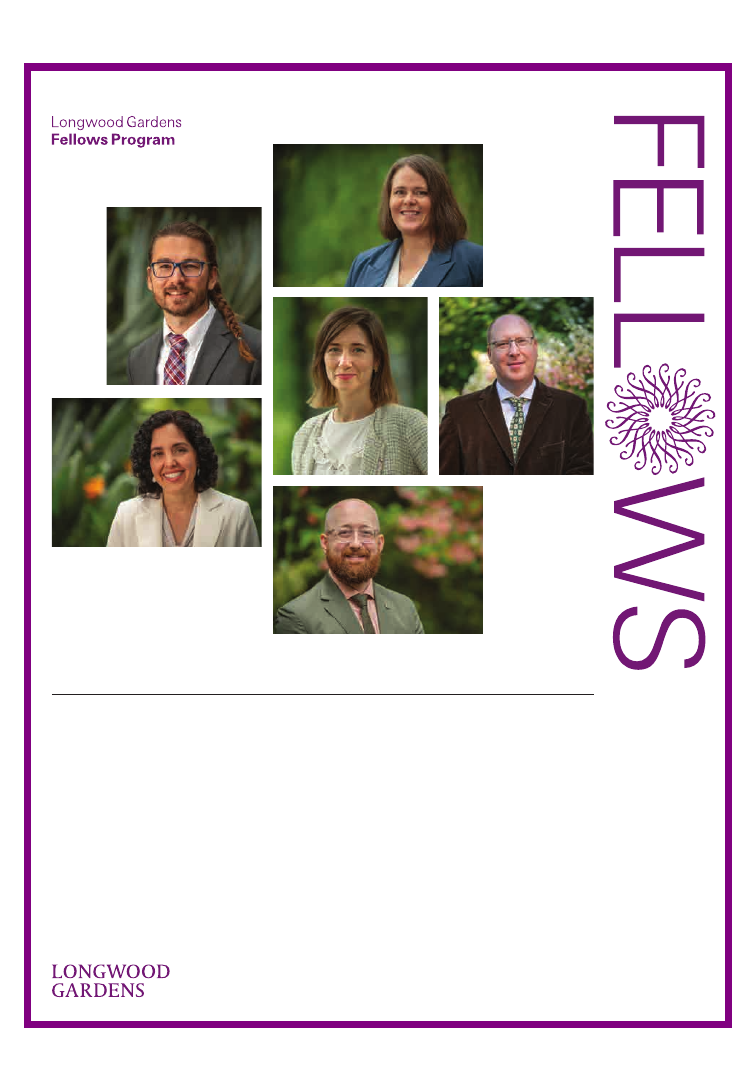
PSB 69 (3) 2023
253
Do you aspire to lead a
horticultural institution
or business?
Are you passionate about
using your career to make
a positive global impact?
Your Path
to Leadership
Applications for the 2024–2025 cohort are
open through July 31. Learn more and apply
at longwoodgardens.org/fellows-program.
Congratulations
to our graduating
2022–2023
Longwood Fellows
Cohort. From top
left: Danny Cox,
Amanda Hannah,
Rae Vassar, Rama
Lopez-Rivera,
Anamari Mena, and
Ryan Gott, Ph.D.
The Fellows Program develops tomorrow’s leaders,
preparing them to successfully navigate pressing
challenges, develop thoughtful strategies, and lead
organizations that are equitable and sustainable.
During the fully funded, cohort-based residency,
Fellows engage in project-based learning that
allows them to hone their professional skills while
delving into issues relevant to the horticulture
industry today.
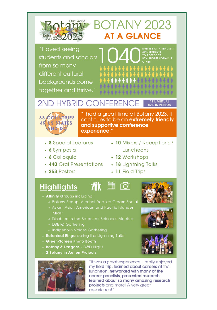
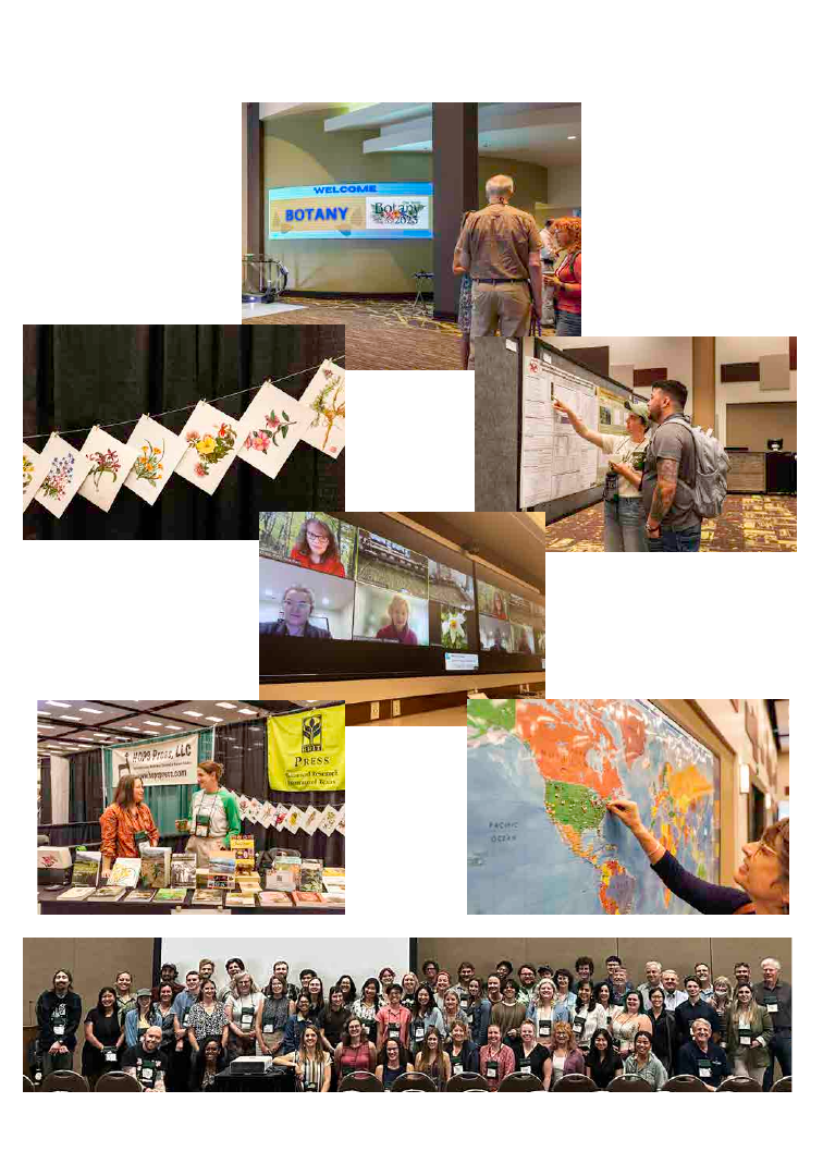
Some Scenes from Botany 2023!
I had a great time at
Botany 2023. It con-
tinues to be an ex-
tremely friendly and
supportive conference
experience.
This was the first
"normal" Botany con-
ference since COVID
and
I liked the energy
people had.
It was nearly back to "nor-
mal"
and, boy, did I need that.
By far, Botany remains one
of the best scientific meet-
ings I attend regularly
California Botanists Brown Bag Lunch!
I thought this was
a great confer-
ence. There were
tons of interest-
ing talks, and the
poster session
was well attended.
Thank you for all
the coffeeeee!!!!
I perceived the conference as a welcoming and
respectful environment where everybody had a
chance to participate and discuss science. I loved
seeing students and scholars from so many dif-
ferent cultural backgrounds come together and
thrive. I especially liked the breakfast time and the
celebration at the end, they were wonderful oppor-
tunities to connect with colleagues and meet new
In grad school I a
ttended
another scientific conference
and went back for the first
time this year - I realized
how much more enjoyable
Botany is and that will be
my main plant conference
in future years. Keep do-
ing what you're doing, it's a
wonderful community!
PSB 69 (3) 2023
257
257
By
Amelia Neely
BSA Membership &
Communications
Manager
E-mail: ANeely@</i>
botany.org
MEMBERSHIP NEWS
The BSA Spotlight Series highlights early-career scientists in the BSA community and shares
both scientific goals and achievements, as well as personal interests of the botanical scientists,
so you can get to know your BSA community better.
Here are the latest Spotlights at https://botany.org/home/careers-jobs/careers-in-botany/bsa-
spotlight-series.html:
• Trinity Depatie, Graduate Student, University of South Carolina
• Adriana I. Hernandez, Postdoctoral Fellow, California Academy of Sciences
• Matias Köhler, Postdoctoral Fellow, São Carlos University (UFSCar), São Paulo, Brazil
• Jesús Martínez-Gómez, Postdoctoral Fellow, University of California, Berkeley
Would you like to nominate yourself or another early career scientist to be in the Spotlight
Series? Fill out this form: https://forms.gle/vivajCaCaqQrDL648.
BSA Spotlight Series

PSB 69 (3) 2023
258
BSA PROFESSIONAL
HIGHLIGHT SERIES
This year, we are including a BSA Professional
Member Highlights section each month in
the Membership Matters newsletter. If you
would like to be highlighted, email Amelia
Neely at aneely@botany.org.
Keri Maricle
Life Sciences Instructor,
Adjunct, at
Barton Community College
LinkedIn:
https://www.linkedin.com/in/kerimaricle/
Keri is a biologist and educator with
experience teaching K-12 and college-level
courses in the life sciences. Currently, Keri
is working on a psychology degree with
research focused on LGBTQ+ inclusion and
sense of belonging in STEM education.
Dr. Jordan Metzgar
Curator of the Massey Herbarium
(VPI), Department of Biological
Sciences, Virginia Tech
X (formerly Twitter):
https://twitter.com/MasseyHerbarium
Dr. Metzgar received his B.S. in biology from
Cornell University and earned his Ph.D.
from the University of Alaska Fairbanks
for his dissertation on the evolution of the
parsley ferns (Cryptogramma). His current
job involves researching the ecology of
various southeastern U.S. plants and running
an active STEM outreach program in the
local community. He became entranced by
ferns when he was an undergraduate, and
his favorite plant is walking fern (Asplenium
rhizophyllum).

PSB 69 (3) 2023
259
NEW BSA AWARD!
AJ HARRIS GRADUATE
STUDENT RESEARCH
AWARD
We are very excited to share that, thanks to
the generous support of over 70 donors, the
Society has established an endowed fund
to support an annual graduate student
research award in memory of AJ Harris. The
online portal for applications will open in a
few months. Learn more about AJ, see a list of
donors, and read the Memory Wall at https://
botany.org/aj-harris-memorial.html.
We want to show our appreciation to AJ’s
friends and family for helping us fulfill the
goals in our strategic plan by supporting
students and botanical research. To discuss
the ways in which you can support the
Society through endowed gifts, please email
BSA Executive Director Heather Cacanindin
at hcacanindin@botany.org.
BSA STUDENT CHAPTER
UPDATES AND EVENT
Help us to welcome to the new BSA
Student Chapters:
• Botanical Society of St. Cloud State
University
• The Gustavus Botanical Society
This is a reminder to BSA Student Chapters’
officers about the upcoming deadline of
December 31, 2023, for the following items:
• Chapters will need to fill out the
event form at https://bit.ly/3rVSe18
for at least two events for 2023. This
will include a description of the event,
possible photos of the the event plus
captions, the number of people who
attended, and if you would like us to
share your event with the BSA com-
munity.
• BSA Student Chapter Presidents
and Secretary/Treasurers will have
to have a current BSA membership.
This means if your current member-
ship expires in 2023, you will need
to renew it by December 31, 2023.
Student chapter members have a dis-
counted membership costing just $10.
We encourage all student chapter
members to have a BSA member-
ship. Financial aid is available by
emailing aneely@botany.org.
• This is a great time to start thinking
about your leadership for the next
year and to have an election if needed.
Please send any leadership changes
to aneely@botany.org as they become

PSB 69 (3) 2023
260
available. New leadership in the Presi-
dent and Secretary/Treasurer posi-
tions will be required to be a current
BSA member for 2024 by December
31, 2023, or when they are elected if
after that date.
If you want to see the full list of BSA Student
Chapters, or learn how to start a chapter at
your institution, go to https://botany.org/
home/membership/student-chapters.html.
IT IS RENEWAL SEASON!
Thank you for your current BSA
membership support! BSA provides annual
memberships that run from January to
December of each year. October is the start
of our membership renewal season, so if your
membership expires in 2023, you should have
started getting renewal notices since early
October.
Please note the following changes starting
October 1, 2023:
• One-Year Affiliate Memberships will increase
by $5, totaling $55.
• One-Year Student Memberships
will increase by $5, totaling $25.
Three-Year Student Memberships will in-
crease by $10, totaling $60.
• One-Year Student and Developing Nations’ Gift
Memberships will increase by $10, totaling $20.
Three-Year Gift Memberships will increase by
$25, totaling $55.
While renewing, you can also renew your
sectional affiliations, donate to the BSA
endowment, award funds, and section
award funds, purchase gift memberships,
and purchase a print subscription of the
Plant Science Bulletin (see below). If you
are not due to renew this year, we hope you
will consider donating to BSA and giving
gift memberships during the fourth quarter.
To renew your membership, go to https://
crm.botany.org. If you do not need to renew,
please consider donating to BSA funds, or
purchasing gift memberships, go to https://
crm.botany.org.
PLANT SCIENCE BULLETIN
PRINT SUBSCRIPTION
CHANGE
Starting with the Spring 2024 issue of
the PSB, print copies will require a $10/
year subscription that will run from
January to December of each year and
will include the three issues for that year.
Subscriptions are now available for 2024 when
you renew your membership. Members who
do not need to renew will be able to purchase
a subscription by going to https://crm.botany.
org/ and choosing the corresponding menu
option.
YEAR-END GIVING
BSA is proud to provide over $120,000 in
awards and grants to our members every
year. Most of these are funded directly by the
generosity of our members via donations to
specific award funds. We hope that you will
consider making a donation to our many
funds including student, professional, and
sectional award funding when you renew your
membership this year. You can also visit www.
botany.org and click Donate to start giving
right now.

PSB 69 (3) 2023
261
Professional members are given the
opportunity of increasing their annual dues by
$25 in order to support the Graduate Student
Research Award fund. Together with GSRA
donations, over $32,202 in additional funds
were raised for the GSRA in the last fiscal year
and 31 GSRA awards were able to be given in
2023. Thank you to all of our members who
made this possible.
The Endowment Fund and the Unrestricted
Fund both have very important roles in the
stability and longevity of BSA. We hope you
will consider making donations to these funds
when choosing your year-end donation plans.
Donations to these funds are being used to
move BSA into the future, and to support our
global community like never before.
Want an even more lasting way to support
BSA? Consider joining the Legacy Society.
To learn more about the society see our latest
Legacy Society email by going to https://
mailchi.mp/botany.org/bsa-legacy-society-
2022-dr1 or visit our Legacy Society web
page at https://botany.org/home/membership/
the-bsa-legacy-society.html.
2023 BSA GIFT
MEMBERSHIP DRIVE
HELP US GET TO 175
GIFT MEMBERSHIPS!
The 2023 Gift Membership Drive has
begun! This year our goal will be 175 gift
memberships through December 31, 2023!
BSA Gift Memberships are a great way to
introduce students and developing nations’
colleagues to the BSA community. You can
purchase one-year ($20) or three-year ($55) gift
memberships by visiting https://crm.botany.
org and choosing “Give a Gift of Membership”.
Don’t have anyone specific for whom to
purchase a gift membership? Not a problem!
You can put an “X” in the gift membership
recipient fields and we will make sure they
get to those students and developing nations’
colleagues who need financial assistance.
Questions about gift memberships or other
ways to donate? Email Amelia Neely at
aneely@botany.org.
We are giving back! Any gift membership
recipient who starts their membership before
January 31, 2024 will be entered into a
drawing for a free registration for Botany
2024!
NEW BSA AD HOC
COMMITTEES
Thank you to everyone who applied to be on
the Ad Hoc Committee on Climate Change,
Membership Ad Hoc Committee, and the Ad
Hoc Committee for AI and Publications. We
appreciate the time and effort taken by those
who did not get chosen and hope that you
will consider applying for open committee
positions as they become available in the Fall.
To see the new committee members, go
to
https://botany.org/home/governance/
committees-committee-officers.html.
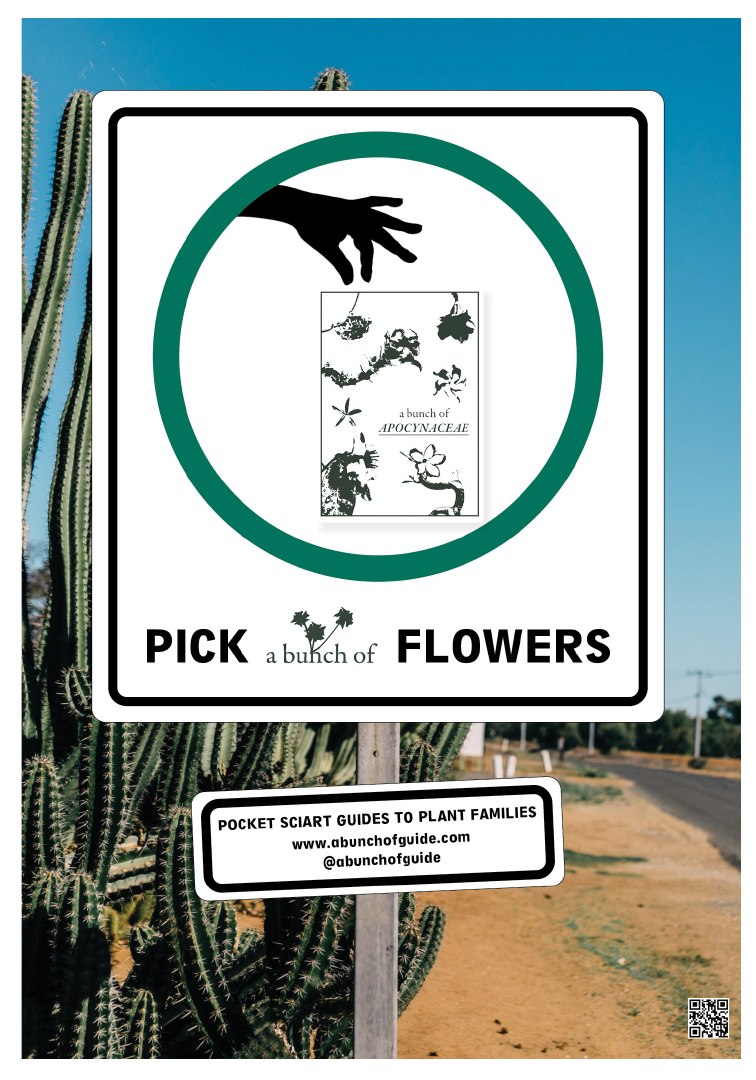
PSB 69 (3) 2023
262

PSB 69 (3) 2023
263
FROM THE
PSB
ARCHIVES
60 years ago
Albert Robinson Jr. from Kansas Wesleyan University discusses research
collaborations with Mexican universities.
“In very recent years an increased emphasis has been placed upon botanical
research in tropical areas. Expanding populations and rapidly developing
industries are placing increased pressure on the existing undisturbed
lands. A sense of urgency has arisen to study and record the biota of
these areas before they are irrevocably altered by man’s quest for a better
life. In this respect, our neighbor to the south, the Republic of Mexico,
offers an excellent opportunity for North American botanists to assist in
this important task, and to work in a tropical region of high botanical
importance which is rapidly being affected by industrialization.”
-Robinson Jr., Albert. 1963. Botany in Mexican Schools. PSB 9(3): 6-7
50 years ago
“At the annual meeting of the Society at Amherst in June, the Council
authorized the establishment of an endowment fund, the income of which
will be used to subsidize the publication of papers in the American Journal
of Botany, and other publications of the Society written and submitted by
students. Immediately upon the establishment of the Endowment Fund a
total of $350 was pledged to the fund from among members of the Council.”
-Endowment Fund Started. 1973 PSB 19(3): 40
40 years ago
“The Teaching Section’s Slide Exchange Program was again very successful
and will be continued and expanded. During the past year over 2500 color
transparencies were duplicated for society members.”
-Teaching Section Slide Exchange Program. 1983. PSB 28(5): 35.

PSB 69 (3) 2023
264
264
SCIENCE EDUCATION
By Dr. Catrina Adams,
Education Director
Jennifer Hartley,
Education Programs
Supervisor
Have you ever had an undergraduate student
or other early-career botanist leave the area(s)
you know best to pursue their botanical
interests in another state? Do you wish you
could easily provide them with the best, most
up-to-date flora or field guide for their new
location? Or have you ever been asked where
someone could find a degree program in
botany or a botanically focused organization
outside of your location?
The BSA Education Committee is seeking
to update the state-by-state resource lists
available on the botany.org website by
crowdsourcing from BSA’s knowledgeable
members. We’re looking for information
about up-to-date floras and field guides,
academic programs (where in your state can
people pursue a botany-related degree?), as
well as organizations and quality, durable web
resources focused on the botany of the state or
region. To start, we are focusing on U.S. states
Updating BSA’s State-by-State
Botanical Resource Pages
Please Help!
and territories, but we may expand this project
to cover other regions where BSA members
live and work if this project is successful and
members find the information useful.
To submit a resource, please use this link:
https://forms.gle/VjpHPYM9pVKJ4dmh9
Together, we can build a valuable reference list
that will help botanists and aspiring botanists
orient to high-quality botanical resources
of states and territories with which they are
not already familiar. It should take less than
5 minutes to submit your resource(s), which
will be vetted by the Education Committee
and then added to the botany.org website.
Thank you very much for your help with this
low-lift but (hopefully) high-value project!
PLANTINGSCIENCE
UPDATES
Summer and Fall of 2023:
F2 Research is Underway
The PlantingScience team has been working
at full tilt this session! With more than 40
teachers signed up between our current
F2 research participants and our returning
teachers from previous years, we are having to
keep more plates spinning than usual.

PSB 69 (3) 2023
265
Fortunately, we had a great summer of
professional development workshops. We
had 21 teachers and 15 scientists take part in
our in-person workshops in Colorado Springs
and St. Louis, and another 18 teachers and
14 scientists participate in online training
via Zoom. These trainings introduced
participants to the activities that comprise
our Power of Sunlight investigation theme,
which focuses on photosynthesis and cellular
respiration, and gave them an opportunity to
experience online “mentoring” as teachers
designed their own exploration with support
from their scientist partners.
In addition, we have 18 teachers who are
acting as a control group this session, teaching
the same topics using their usual curriculum
and activities. These teachers will be included
in workshops during summer of 2024 and will
use PlantingScience with students during the
Fall 2024 session.
MEET OUR 2023
F2 FELLOWS!
Please join us in welcoming and congratulating
the early career scientists who are participating
in the F2 research work this session:
Abdulkabir Abdulmalik
Hannah Assour
Israel Borokini
Meghan Britton
Jessica Carstens-Kass
Snehanjana Chatterjee
Cael Dant
Natalie Dietz
Kasia Dinkeloo
Chloe Fackler
Josh Felton
Melinda Findlater
Julie Gan
Audrey Geise
Kajal Ghoshroy
Devani Jolman
Harkirat Kaur
Janet Mansaray
Deannah Neupert
Wanderson Novais
Sofia Ocampo
Varsha Pathare
Adam Ramsey
Philippa Stone
Jessica Szetela
Nicole Vaccaro
Imeña Valdes
Gabriela Villani
Renate Wuersig

PSB 69 (3) 2023
266
READ ABOUT
PLANTINGSCIENCE
DIGGING DEEPER
RESEARCH RESULTS:
FREE ARTICLE IN THE
AMERICAN BIOLOGY
TEACHER
An article on the efficacy of the PlantingScience
Power of Sunlight program has been selected
as the free article in the September issue of the
American Biology Teacher:
https://nabt.org/
files/galleries/ABT_Online_Sept_2023.pdf
This article reports on the results of our
2015–2019 research studying the efficacy
of the PlantingScience Power of Sunlight
module. The research showed positive gains
in participating students’ content knowledge
and attitudes about scientists over students
who learned photosynthesis and cellular
respiration the way their teachers normally
taught those topics.
We’re honored to be selected and hope that
the article will have a wider reach since it is
open access!
;@TorreyBotanical
www.torreybotanical.org
;@TorreyBotanicalSociety
Torrey Botanical Society
THE OLDEST BOTANICAL SOCIETY IN THE AMERICAS
Field trips
held in the
NY/NJ/CT area
Journal of the Torrey
Botanical Society
free to publish
low open-access fees
Virtual lectures
watch our past
lectures on YouTube
Undergraduate,
graduate, and early
career fellowships
application deadline:
January 15
Since our founding in New York City in 1867, the
goals of the Torrey Botanical Society have
remained the same: to promote an interest in
botany, and to collect and diffuse information
on all topics relating to botany.
Staten Island, 1914
upstate NY, 2012
PSB 69 (3) 2023
267
Plant Conservation &
Restoration Program
U.S. Department of the Interior
Bureau of Land Management
Native plants are the true green infrastructure we rely on
for healthy, resilient, biodiverse ecosystems. As
wildfires and other climate-driven disasters continue to
devastate the U.S., the BLM Plant Conservation &
Restoration Program is implementing the National Seed
Strategy and conserving and restoring the native plant
communities that define America's iconic landscapes
and provide wildlife habitat, ecosystem services, and
recreational opportunities for all Americans to enjoy.
blm.gov/nativeplants
blm.gov/seedstrategy

PSB 69 (3) 2023
268
268
STUDENT SECTION
By Eli Hartung and Josh Felton
BSA Student Representatives
It was so great seeing so many of your faces
again in person at Botany 2023 in Boise!
Students made up 53% of the total conference
attendees with 494 total students. About a
quarter of the students were undergraduates
and three quarters were graduate students.
About 85% of total students attended the
conference in person. The popularity of face-
to-face attendance at Boise highlighted how
important it is to have in-person interactions
to cultivate our botanical network. At the
same time, about 15% of students attended
virtually. We hope the opportunity to tune
in remotely helped students stay connected
to the botanical community this year, even
though they couldn’t be there in person.
The Student Reps worked to encourage
more interactions between students and the
botanical community at a variety of events.
Our first event of the week was the Writing
Your CV and Translating it Into a Public Facing
Website on Sunday, where seven superstars in
their respective areas of sci-comm shared tips
for engaging audiences about plants. The next
day, we held the widely popular Careers in
Botany 2023 Review
Botany Luncheon where we had 12 panelists
for students to connect with. We then helped
host a very well–attended Student Social at
the Linen Building where we chatted late
into the night. We also held a virtual Student
Chapter Meetup where we discussed ideas to
connect members with other chapters across
the country.
Reach us by email or X (formerly Twitter):
Eli at elishartung@gmail.com / @hartung_
eli or Josh at feltonjosh@icloud.com /
;@JoshFelton12.
CAREERS IN BOTANY
LUNCHEON
At the Careers in Botany Luncheon,
we had 12 panelists with careers in
academia, government, non-governmental
organizations, consulting companies,
herbaria, botanical gardens, and museums.
They represented the spectrum of career
stages, and collectively, they work in five
countries and ten U.S. states. A total of 106
students attended—one of our best attended
luncheons yet! Below was the flier we used to
advertise the event, and here is where you can
read more about the panelists at the Careers
in Botany Profiles: https://botany.org/home/
careers-jobs/careers-in-botany/careers-in-
botany-profiles-2023.html

PSB 69 (3) 2023
269
STUDENT SOCIAL
Thank you to the 143 of you who attended the
Student Social! We had a great time getting
to know each other at the Linen Building in
Boise. Hope to see you all again at the next
student social in Grand Rapids, Michigan!
WRITING YOUR CV AND
TRANSLATING IT INTO A
PUBLIC FACING WEBSITE
WORKSHOP
Our first CV writing/website building
workshop was a great success with 7 panelists
and 12 attendees. The workshop panelists
were a diverse group of plant scientists with
experience in both CV building and website
management. First, the panelists spent a few
minutes introducing themselves and their
work. Then, panelists presented tips on CV
building/formatting and provided feedback
to students on their CVs. Finally, panelists
showed their websites and provided advice as
students built their own websites. We learned
so much from the panelists, both through
their engaging presentation content and style,
and in the super interesting small discussions.
Read the section “Heard at the Writing Your
CV and Translating it Into a Public Facing
Website Workshop” to learn some tips from
our panelists.
STUDENT CHAPTER
MEET-UP
We held our first ever Virtual Student Chapter
Meet-up at the conference this year. The event
was initiated and organized by our very active
student chapters. We would love to host this
at the conference each year. To maintain this
momentum, the Student Reps are planning
more of these Student Chapter Meet-ups as
part of the Botany360 program. Stay tuned on
the @Botanical_ X (Twitter) and BSA Student
Newsletter to hear more details.
NETWORKING BOARD
For our conference Networking Board, we
heard from 7 labs recruiting for more than 13
positions including Masters and PhD student,
research assistant, post-doc, and technician
openings in 7 states across the country. Be
sure to reach out to them using the contact
information listed on the board!
We also heard from 18 early-career
researchers looking for graduate school
positions, post-docs, jobs in industry,
government, and lab or field positions. Those
of you recruiting, please check out this list!
https://tinyurl.com/networkingboard
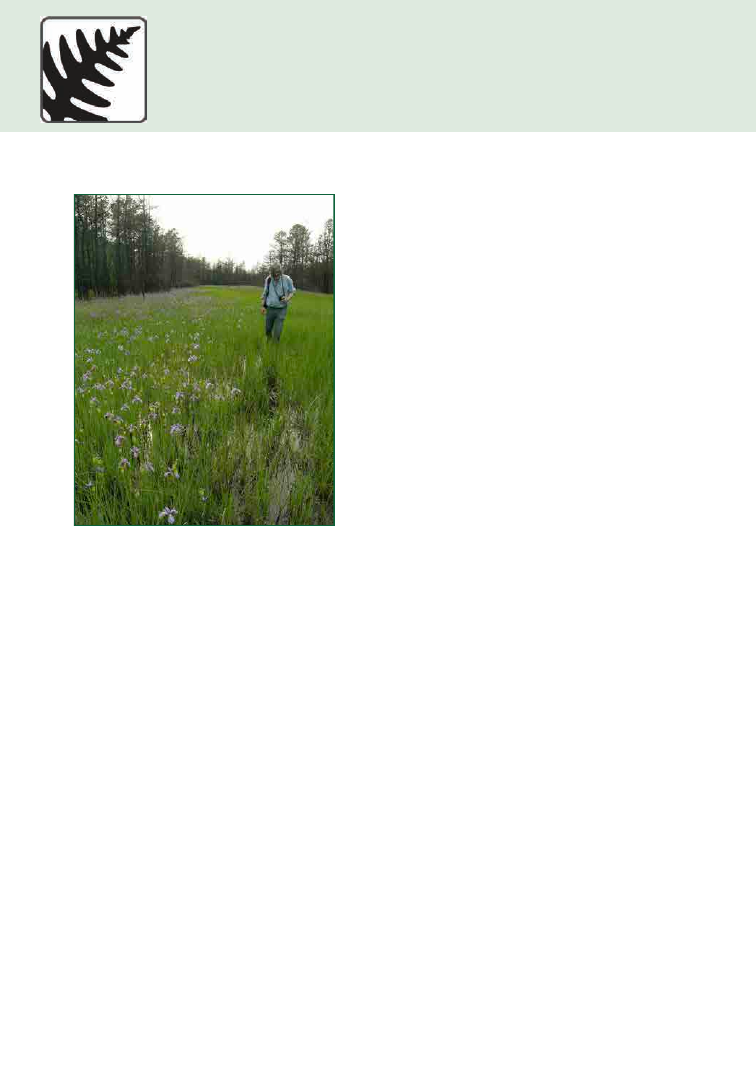
PSB 69 (3) 2023
270
270
ANNOUNCEMENTS
IN MEMORIAM
JOEL FRY
(1957-2023)
It is with great sadness that we note the
passing of Joel Fry on the 21 March 2023.
Joel was a world-renowned scholar centered
at Bartram’s Garden, America’s oldest
surviving botanic garden, founded in 1728
by John Bartram and still a working historical
garden on the western bank of the Schuylkill
River running through Philadelphia. Joel’s
work at Bartram’s Garden encompassed
archival history, archaeology, anthropology,
horticulture, gardening, field botany, and so
much more. Joel generously shared his wealth
of knowledge with scholars, amateurs, and all
who showed an interest in the world around
us. He reached across disciplines and counted
historians and gardeners and botanists among
his numerous friends and colleagues.
Ph
ot
o p
ro
vi
de
d b
y A
llen C
ra
w
fo
rd.
As just one example of Joel’s ability to work
across disciplines, during digitization of
specimens at the Herbarium of the Academy
of Natural Sciences (PH), a specimen collected
shortly after the Battle of Gettysburg was
discovered. The specimen was collected by
Thomas Meehan who, in addition to being a
member of the Academy of Natural Sciences
of Philadelphia (Botany Department) and
a prominent nurseryman in Philadelphia,
had worked at Bartram’s Garden early in his
career, shortly after he arrived in the U.S. from
England. This connection led to a scholarly
partnership that Joel dove into, providing
numerous and eloquently written inputs,
which led to a publication in 2022 (McCourt
et al., 2022).
Joel got his MS in Historical Archeology from
the University of Pennsylvania and began
working at Bartram’s Garden in 1992. He
worked there for more than 30 years until his
death at age 66. Joel always welcomed visitors
to Bartram’s Garden, including neighbors and
people visiting from near and far, including
numerous visitors from all over the world;
students from local colleges and universities;
and members of local natural history
organizations like the Philadelphia Botanical
Club and the Delaware Valley Ornithological
Club.
The first field trip of the Philadelphia Botanical
Club was to Bartram’s Garden, in 1892, and
in 2012, to celebrate the 120
th
anniversary
of that event, Joel led a botanical excursion
at Bartram’s. Only three species were listed
from that 1890s field trip report (Eranthis
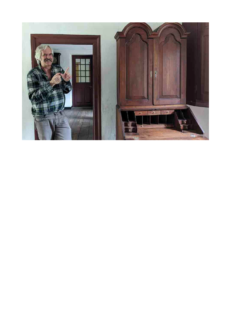
PSB 69 (3) 2023
271
hyemalis [winter aconite], Ptelea trifoliata,
and Aralia spinosa). Joel assiduously researched
where those species might and might not
be at Bartram’s, and only one, E. hyemalis,
is still found at Bartram’s Garden (https://
growinghistory.wordpress.com/2012/02/24/
a-visit-to-bartrams-garden-with-the-
philadelphia-botanical-club/). This is an
example of the diligence and thoughtfulness
that Joel put into all his work. Joel’s scholarly
publications are nonpareil, and his work
outside of academic publishing equaled the
excellence of his more formal scholarly work.
However, the thought of calling anything
having to do with Joel “formal” would seem
outlandish to all of us who knew him—Joel’s
lack of formality undergirded everything he
did. If he could learn something and share it
with the world, he did all he could to do that
work with excellence.
Joel traced the movement of plants across the
Atlantic (Fry, 1996) and throughout North
America (Fry, 2000). Joel’s rigorous research
on the archival history and the botany of plant
movement leads to knowledge that critically
informs the biology of the movement of plants,
in cultivation, as well as naturalization, and
increases our understanding of how extinction
in the wild interacts with horticulture and
conservation in gardens.
Joel contributed freely to a range of scholars,
as a glance through the acknowledgments
of numerous papers clearly shows (e.g.,
Schoonderwoerd and Friedman, 2016;
Gladfelter et al., 2020). Acknowledgment of
Joel’s contributions in many books also shows
his generosity of spirit (for example, Andrea
Wulf’s Founding Gardeners; and Victoria
Johnson’s American Eden).
Joel Fry showing off the newly acquired Bartram desk in the Historic Bartram House in
Summer 2022. Photo provided by Bartram’s Garden.

PSB 69 (3) 2023
272
One of Joel’s unfinished projects was a study of
southern New Jersey plants the Bartrams knew.
This began with his and Bill Cahill’s realization
that the Sutro Herbarium at the Huntington
Library contained specimens John Bartram
must have collected in “Jersey,” because this
hortus siccus predated his trips further south
than Delaware (then part of Pennsylvania)
and Maryland. They recognized, for example,
the Diapensia species Pyxidanthera barbulata
from a shriveled, disintegrating specimen,
and the pine barren gentian Gentiana
autumnalis, which Bartram couldn’t have
found closer to home. With this they began
to look further, eventually listing a few
hundred plants from the Sloane Herbarium
in London (once it became available in digital
online images) and other sources that gave
a detailed understanding of the Bartrams’
local botanical knowledge. John and William
Bartram were famous botanical travelers, but
the evidence of this study showed that they
learned much of their botany locally, even as a
preparatory study for understanding plants in
the far-off regions they visited. Fry and Cahill
also traced reiterations of this botanic interest
in the flora of Philadelphia and southern New
Jersey in the work of William P.C. Barton
(1818), Nathaniel Lord Britton (1880), and
Ida Keller and Stewardson Brown (1904).
They also found Bartram provenances for
taxa named by others, such as the pine barren
shrub Leiophyllum buxifolium Bergius, which
was communicated to Bergius by a Swedish
correspondent who knew the Bartrams in
Philadelphia.
On the 7th of September 2023, a gathering
was held at Bartram’s Garden in Philadelphia
in remembrance of Joel, in a place that not
only was a center for him, but in many ways,
one might say, was centered on him. Many
gathered and remembered his knowledge
(phenomenal), his erudition (brilliant), his
sense of humor (high spirited), his generosity
(endless), and most of all, his friendship,
which will be dearly missed.
REFERENCES
Fry, J. T. 1996. An international catalogue of
North American trees and shrubs: the Bartram
broadside, 1783. Journal of Garden History
16: 1: 3-66.
Fry, J. T. 2000. Franklinia alatamaha, A His-
tory of That “Very Curious” Shrub. In: Bar-
tram Broadside, Special Franklinia Edition,
published by the John Bartram Association
for the ‘noble & curious friends’ of Historic
Bartram’s Garden.
Gladfelter, H. J., L. K. Yadav, S. A. Merkle,
et al. 2020. Genetic diversity and population
structure analysis of Franklinia alatamaha, a
tree species existing only in cultivation. Tree
Genetics & Genomes 16: 60.
McCourt, R. M., J. T. Fry, and E. Benamy.
2022. A Flower from Gettysburg. Bartonia
71: 1–8.
Schoonderwoerd, K. M., and W. E. Friedman.
2016. Zygotic dormancy underlies prolonged
seed development in Franklinia alatamaha
(Theaceae): a most unusual case of reproduc-
tive phenology in angiosperms. Botanical
Journal of the Linnean Society 181: 70–83.
--By David Hewitt, Richard McCourt, and
William Cahill
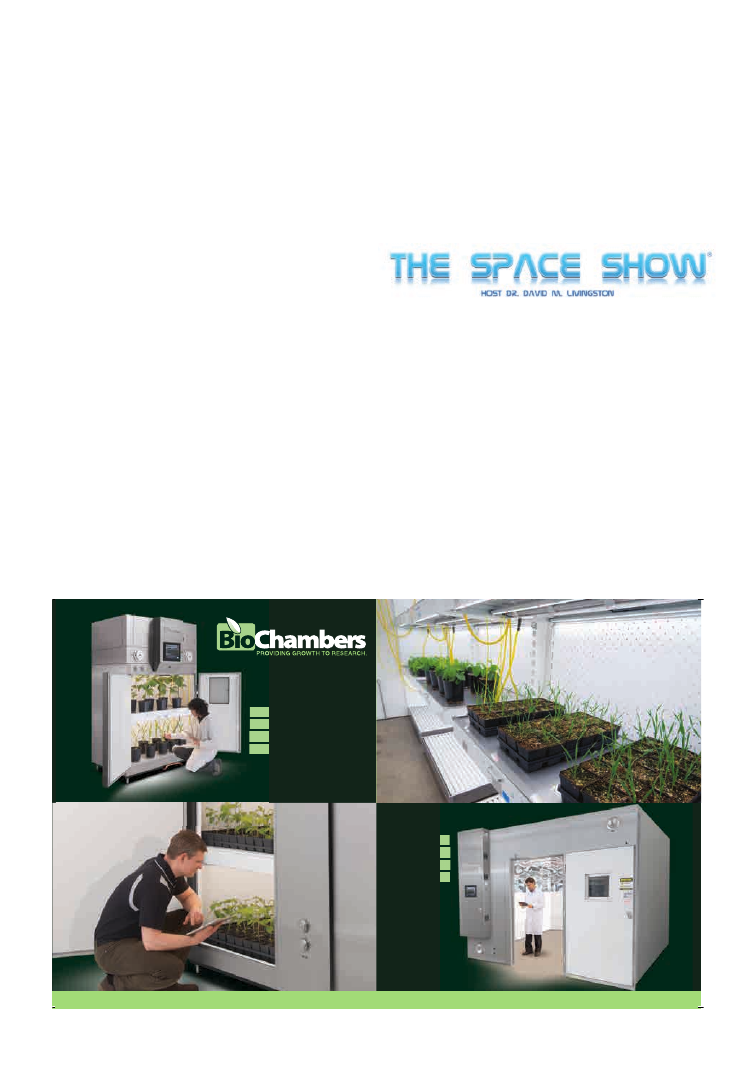
Walk-in
Plant
Growth
Rooms
Reach-in
Plant
Growth
Chambers
biochambers.com
477 Jarvis Ave, Winnipeg, Canada, R2W 3A8
Toll Free: 1-800-361-7778
DR. JOHN KISS
FEATURED ON THE
SPACE SHOW
BSA Member John Kiss was recently featured
on an episode of the radio show The Space
Show. Topics included gravitational and
space plant biology for the Moon, Mars,
free space, Cislunar space, and more. Kiss
also talked about plant needs, challenges,
human components, radiation, the role of
microgravity, and the quality of light plus
water needed for space biology.
The episode can be found at: https://www.
thespaceshow.com/show/18-aug-2023/broad-
cast-4075-dr.-john-z.-kiss.
PSB 69 (3) 2023
274
274
A Generic Classification of the Thelypteridaceae
Flora of Colorado, Ed. 2
Getting In: The Essential Guide to Finding a STEMM Undergrad Research
Experience. 2
nd
Edition
Jackfruit: Botany, Production and Uses
Mistletoes of the Continental United States and Canada
Moving Crops and the Scales of History
Orchid
Planting Clues: How Plants Solve Crimes
Putting Down Roots: Foundations of Botany at Carolina with a Concluding Chapter on the
History of the Department of Botany
Stelar Evolution and Morphology In Selected Taxa Based On The Study Of Vascullotaxy
(Studio Nov.), Ed 2
Taylor’s Seedling Drawings: A Catalog of Cotyledons
BOOK REVIEWS
A Generic Classification of
the Thelypteridaceae
Susan Fawcett and Alan R. Smith
2021. ISBN 13: 978-1-889878-68-3
Flexbinding, US$25.00; 112 pp.
BRIT Press
Although presented as a book,
this work is more like a long
journal paper covering the classification of
the Thelypteridaceae. It is laid out just like
any other such paper with an introduction,
illustrations, keys, etc. The classification of the
fern genera reflects phylogenetic work done
by the authors that was also published in 2021.
Many gardeners bemoan the name changes
that angiosperms have undergone in the last
few decades, but if only they knew what has
happened with fern classification! The advent
of next-gen sequencing has given us a much
deeper insight into the evolutionary history
of these fascinating plants; therefore, many
new names have been created and old ones
reinstated. Fawcett and Smith’s work here
clarifies this particularly complicated family
and provides a new classification that was
sorely needed. The authors publish 176 new
names and recognizes 37 genera. The authors
do a good job reviewing the taxonomic
history of the family and the botanists that
came before them who tried to make sense of
these plants. A number of line drawings and
photos are included to illustrate the ferns. The
bulk of the paper consists of the taxonomic
descriptions of each genus, which also include
the history of each one’s circumscription,
geography, and references.
The wealth of information in this work is
wonderful and it is apparent that the authors
truly enjoy working with these plants. Anyone
looking to know more about the family
or put updated and correct names to their
Thelypteridaceae specimens should purchase
this.
-John G. Zaborsky, Botany Department,
University of Wisconsin – Madison, Madison,
Wisconsin, USA; jzaborsky@wisc.edu

PSB 69 (3) 2023
275
Flora of Colorado, (ed. 2)
Jennifer Ackerfield
2022. ISBN 978-1889878898
US$80; 872 pp.
BRIT Press
Colorado’s flora is rich due
to the diversity of habitats,
from the Eastern Plains to
the Rocky Mountains and the high deserts
of the Western Slope. To help botanists
identify the plant species, there have been
numerous floristic works, from the checklist
of Porter and Coulter’s “Synopsis of the Flora
of Colorado” (1874) to the seminal “Manual
of the Plants of Colorado” (1964) by Harold
Harrington. More recently, William Weber
and Ronald Wittmann divided the state
at the Continental Divide and published
separate floras for the Eastern and Western
Slopes, with the fourth editions published in
2012. These floras contained keys but lacked
species descriptions and distribution maps,
and often used unconventional nomenclature.
For many users, this last feature made Weber
and Wittmann’s floras difficult to use. As a
resident of western Colorado, I appreciated
a flora that focused on plants of my region
while excluding those of the eastern plains.
However, a modern flora covering the entire
state was lacking.
In 2015, Jennifer Ackerfield published the first
edition of the Flora of Colorado. This up-to-
date flora used conventional nomenclature,
taxonomic changes ushered in by the
molecular age, succinct species descriptions,
distribution maps, and 912 images covering
nearly one third of the plant species in the
state. I appreciated the county map on inside
of the front cover and the ruler on the inside
of the back cover, an indispensable feature for
field identification. To say the first edition
was well-received by Colorado botanists is an
understatement! It quickly became the go-to
flora for Colorado botanists.
The second edition of the Flora of Colorado
includes many updates. Nomenclatural
changes have been included as have new
taxa that have been recognized, collected,
or identified since the first edition. The first
edition contained 3322 taxa (including 645
varieties and subspecies) whereas the second
edition contains 3352 taxa. Among the new
taxa are two species of Cirsium described by
Ackerfield in 2022.
The second edition is beautiful, with a cover
photo of a misty mountain meadow with
Veratrum and Delphinium. The cover of the
second edition seems to be made of thicker,
tough paper then the first edition. This is a
welcome addition, given how torn and tattered
my well-used first edition has become. The
second edition is a full centimeter thinner
than my first edition due to the slightly wider
pages. It is still a rather large volume to take
into the field, but the ability to key out any
plant in Colorado is well worth the extra
weight in a backpack.
New distribution maps are one of the major
and most eye-catching updates. The first
edition included county-level distribution
maps. These provided a rapid overview of
distribution, but the new dot-distribution
maps give more detail as to exactly where
collections have been made as well as a
general idea of how frequently specimens
are collected. These improvements are due to
large-scale databasing efforts and availability
of specimens online. It took a little time for
me to get used to the dot-distribution maps
and the dots are small, making them a bit
difficult to see; however, I appreciate the added
precision. The discussion of the maps as well
as the heat map of herbarium collections (p.
14) are quite interesting, and I appreciate
that Ackerfield takes the time to discuss their
creation, what data were included, and what
data were excluded.

PSB 69 (3) 2023
276
The images in the second edition are greatly
improved. There are 1296 color images on 108
plates (versus 912 images in the first volume).
Not only is the quantity improved, but these
images are more informative. I particularly
appreciated the inclusion of multiple photos,
such as in the Cyperaceae and Poaceae plates,
covering both fine-scale characters like the
flowers as well as overall appearance of the
inflorescences. Additionally, Ackerfield has
included black-and-white images within the
keys for particularly difficult taxa. The images
of Amaranthaceae bracts and fruits (pp. 78-
79), Boraginaceae nutlets (pp. 230-231), and
Nyctaginaceae anthocarps (pp. 575-578)
are all helpful when looking at difficult but
diagnostic characteristics.
The keys in the first edition were easy to use,
generally included many characters, and
worked well. My initial impression is that the
keys have not changed significantly, although
with more use differences may come to light.
Given how thorough and well done the first
edition was, it is not surprising that there are
not significant differences.
Compiling a flora will always come with
compromises. This is a technical key, so those
wanting a plant identifier picture book should
look elsewhere. One criticism of the Flora of
Colorado is that the species descriptions are
not complete. In my opinion, between the
description of the genus, the key highlighting
the difference between taxa, and the succinct
descriptions, the Flora of Colorado does an
excellent job giving a description of taxa
without redundant, unnecessary verbiage.
Shorter descriptions are also necessary to
keep the length and weight of the book down
so that it can be taken into the field.
While I will keep hold of my torn and tattered
first edition, I am thrilled to have a new,
updated edition of the Flora of Colorado. The
first edition of the Flora of Colorado was a
huge leap forward for Colorado botanists and
the second edition finetunes this work. The
value of this work cannot be underestimated.
I experienced this firsthand during my Plant
Identification course in the Fall of 2022 when
the first edition was out of stock and the second
edition had yet to be released. I did not fully
appreciate the value of this flora until it was
unavailable! Botanists in the state are lucky to
have such a thorough yet concise flora.
REFERENCES
Ackerfield, J. 2015. Flora of Colorado, ed 1. Bot. Misc.
41. BRIT Press, Fort Worth Botanic Garden, Botanical
Research Institute of Texas, USA.
Ackerfield, J. 2022. Flora of Colorado, ed 2. Bot. Misc.
60. BRIT Press, Fort Worth Botanic Garden, Botanical
Research Institute of Texas, USA.
Harrington, H. D. 1964. Manual of the Plants of Colo-
rado, ed 2. The Swallow Press, Chicago, Illinois, USA.
Porter, T. C., and J. M. Merle. 1874. Synopsis of the Flora
of Colorado. Washington DC, Govt. Publication. DOI:
https://doi.org/10.5962/bhl.title.46971
Weber, W. A., R. C. Wittmann. 2012. Colorado Flora,
Western Slope, ed 4. University Press of Colorado,
Denver, Colorado, USA.
--Stephen Stern, Ph.D., Professor of Biology,
Department of Biological Sciences, Colorado
Mesa University, Grand Junction, CO 81501;
Email: sstern@coloradomesa.edu

PSB 69 (3) 2023
277
Getting In: The Essential
Guide to Finding a STEMM
Undergrad Research Expe-
rience. (ed 2)
Paris H. Grey and David G.
Oppenheimer
2023. ISBN-10: 0226825418;
ISBN-13: 978-0226825410
US$20.00 (soft cover); 253 pp.
University of Chicago Press
Undergraduate research is a high-impact
practice that can transform the lives of students
and lead to increased retention of students
(Weber and Myrick, 2018; Lanning and Brown,
2019). This type of experience certainly made
a big impact on my life and is the reason I am
an academic today. As an undergraduate, I did
two different independent research projects—
one in field ecology and another in electron
microscopy—and then decided that graduate
school and academia was for me. Of course,
undergraduate research in STEM fields can
be for almost all students and can become a
valuable part of their training.
As a faculty member, I have come full
circle and continue to enjoy mentoring
undergraduates in independent research.
Some of these students go to graduate school
and others to medical school. In the latter case,
I always think that it is good for physicians to
understand the nature of scientific research.
My best students have been co-authors on
papers. One of my undergraduates (who is
now a tenured faculty member) discovered
a novel photosensory mechanism in plants
(Ruppel et al., 2001), which served as the basis
for a successful grant proposal for a spaceflight
project with NASA (Millar et al., 2010; Kiss et
al., 2012)!
This book provides a fantastic and
comprehensive overview of all aspects of
undergraduate research in STEM fields. Part
1 (Chapters 1–3) covers the basics such as
the benefits of research as an undergraduate,
a primer on types of STEM research, and an
overview of lab culture. Part 2 (Chapters 4–6)
considers detailed strategies of applying for
research positions as an undergraduate.
There are many reasons to pursue
undergraduate research, and a few are
outlined above. However, Chapter 1 provides
a very thorough list of the benefits—both
obvious and less-than-obvious ones. For
instance, it is well known that undergraduate
research will help get a student into graduate
school. Other benefits and skills include:
upholding a commitment, working well with
others, embracing constructive feedback,
developing organizational strategies,
developing outstanding communication
skills, strengthening self-discipline, learning
time-management skills, improving critical
thinking, among others. The authors speak
of these benefits with conviction and passion.
Chapter 2 is an introduction to research
groups and lab culture in STEM and serves
as a good overview of the research process.
The authors consider lab work, field research,
clinical studies, and other types of research.
The authors also posit in Chapter 3 that
undergraduate research is great for all types
of students (Laursen et al., 2010)—not
only honors or high-GPA students—and I
completely agree. To make this point, the
authors write, “Most scientists aren’t geniuses
or even brilliant. What they are is interested
in their research and resilient when managing
disappointment and failure.” This last
statement is one of the most accurate ones that
I have read about scientists and gives insight
into the nature of science. The answer to the
question of when a student should start a
research project is given by the comment that
PSB 69 (3) 2023
278
you should start your research experience as
soon as you can handle the time commitment.
The advantage of starting sooner is that the
longer you are in the lab, the more likely
that you will have a meaningful research
experience (e.g., attend scientific meetings,
perhaps co-author a paper).
The next several chapters (Part 2 of the book)
cover all aspects of applying for research
positions. One consideration is whether the
student wants to do work at their own college
or university or apply to one of the many
Summer Undergraduate Research Programs
(SURPs). This book is very comprehensive
in all aspects, and the authors consider items
such as application strategies and how to
interview with a Principal Investigator.
The authors are both experienced science
faculty who have mentored many students
in their labs. They also actively promote
undergraduate research via social media
such as Instagram and X (formerly Twitter):
;@YouInTheLab. This book is great for many
groups such as undergraduates thinking about
research or those already engaged in such
work as well as their faculty mentors. It can be
used as part of a course that meets to provide
mentoring for undergraduate researchers and is
an inexpensive, enjoyable, and a fun read as well.
REFERENCES
Kiss, J. Z., K. D. L. Millar, and R. E. Edelmann. 2012.
Phototropism of Arabidopsis thaliana in microgravity
and fractional gravity on the International Space Sta-
tion. Planta 236: 635–645.
Lanning, S., and M. Brown. 2019. Undergraduate re-
search as a high impact practice in higher education.
Education Sciences 9: 160.
Laursen, S., A. B. Hunter, E. Seymour, H. Thiry, and G.
Melton. 2010. Undergraduate research in the sciences:
Engaging students in real science. John Wiley & Sons.
320 pp.
Millar, K. D., P. Kumar, M. J. Correll, J. L. Mullen, R.
P. Hangarter, R. E. Edelmann, and J. Z. Kiss. 2010. A
novel phototropic response to red light is revealed in
microgravity. New Phytologist 186: 648-656.
Ruppel, N. J., R. P. Hangarter, and J. Z. Kiss. 2001.
Red-light-induced positive phototropism in Arabidop-
sis roots. Planta 212: 424-430.
Weber, K., and K. Myrick. 2018. Reflecting on reflect-
ing: rummer undergraduate research students’ experi-
ences in developing electronic portfolios, a meta-high
impact practice. International Journal of ePortfolio 8:
13-25.
--John Z. Kiss, Department of Biology, UNC-
Greensboro, Greensboro NC 27402
Jackfruit: Botany,
Production and Uses
Sisir Mitra, Editor
2023. ISBN: 978-1800622296
US$170 (Hardcover); 292 pp.
CABI Publishing, Oxfordshire,
U.K.
Jackfruit: Botany, Production and Uses
comprises 13 chapters dedicated to exploring
the life and times of jackfruit, the fruit-bearing
evergreen tree. It begins with a botanical
introduction of the Moraceae family, of which
jackfruit (Artocarpus heterophyllus), figs,
and breadfruit are members. Mitra’s Preface
provides readers with a brief account of
jackfruit’s origins, noting that it is native to the
Western Ghats mountain range, which runs
north–south along the western coast of India.
Because of the tropical climate of the lowlands
in South and Southeast Asia, where jackfruit
is cultivated, it enjoys a long growing season.
Jackfruit has several purposes, providing
major jackfruit-growing countries such as
Bangladesh, the Philippines, and Vietnam
with food, fuel, timber, and medicinal and
industrial products (p. xi). Jackfruit is also
grown in Belize, Jamaica, and Florida. And
although jackfruit is a major staple in South

PSB 69 (3) 2023
279
and Southeast Asia, it is an understudied plant,
taking a backseat to the avocado, mango, and
coconut tree.
Most things a horticulturalist, botanist,
farmer, and crop scientist may want to know
about jackfruit cultivation, including breeding
and propagating methods, can be found in
Jackfruit. There are directions for propagating
jackfruit with cuttings or by grafting. There is
information about soil nutrition, fertilization,
and irrigation practices for increasing the
quality of the tree and its fruit. Orchard
managers will find the “Orchard Management”
section instructive as it provides a roadmap
for starting and maintaining a jackfruit
plantation, replete with information about
pruning and training and intercropping.
Importantly, while jackfruit is easy to grow
and maintain, the “Biotechnology” chapter
stresses the importance of using such tools as
plant tissue culture, genetic engineering, and
marker-assisted selection to improve crop
production.
The very climate in which jackfruit thrives
also invites such pesky pests as the jackfruit
borer, which burrows into the fruit and shoot
of the tree, and the red-spotted longhorn
beetle, which can wreak havoc on jackfruit’s
stem and trunk system. Fruit flies and bark-
eating caterpillars, which also feast on the
fruit and bark of the tree, are also a nuisance
for the jackfruit. Plant physiologists, forest
entomologists, and forest pathologists whose
research focus is on the pests, diseases,
and physiological disorders afflicting the
evergreen will find these chapters valuable.
Each provides specific details on the lifecycle
of the pests and diseases; the specific trajectory
they follow in young and mature jackfruit tree;
their effects on the whole tree, from crown
to root; and what pest and disease control
techniques can be employed to ensure strong
growing conditions that produce high-quality
jackfruit trees.
Although jackfruit trees are not free from pests
and disease, they are termite resistant, which
is why the tree’s beautiful golden-colored
wood is used to build furniture, houses, and
boats. In India, the timber is used to build
the veena, a string instrument resembling a
sitar (Chandrashekhar et al., 2021). Jackfruit
also grows naturally in backyards and is
used as a windbreaker, lining major avenues
in countries such as Bangladesh and Sri
Lanka. In addition to being drought resistant
(jackfruit can tolerate about 3 to 4 months of
dry period, and it can tolerate a bit of frost),
it can withstand strong winds and has been
shown to survive hurricanes, recovering well
from losing leaves and branches (Elevitch and
Manner, 2006, pp. 6–7).
Those herbalists and ethnopharmacologists
interested in the medicinal and
pharmacological attributes of jackfruit
will find Bhattacherjee’s “Composition and
Uses” chapter a useful introduction. As a
nutraceutical, jackfruit has many medicinal,
nutritional, and health-giving benefits,
and Bhattacherjee notes that the bioactive
compounds flavonoid and phenolics in
jackfruit serve as powerful antioxidants, anti-
inflammatory, and anti-cancer agents and
have been used to successfully treat cancers of
the skin, lung, and breast (p. 40). He also notes
that jackfruit is “one of the rare fruit sources
of B Vitamins” (p. 40).
One of the best features of Jackfruit is the
“Processing and Products” chapter, which
explores how the whole tree is processed and
what kind of food and industrial products it
yields. When it is green and unripe, jackfruit
can be cooked and eaten as a vegetable, and
when it is ripe, jackfruit is eaten as a fruit.

PSB 69 (3) 2023
280
Its seeds can also be boiled and sauteed
or roasted. One can enjoy jackfruit candy,
jackfruit chips, and jackfruit ice cream. Chefs
who are interested in cooking the rice dish
biriyani, for instance, will find step-by-step
instructions. The tree is so versatile that it
is even used as a meat substitute; its fibrous
texture resembles meats like chicken or pork
(Ghangale et al., 2022).
Overall, Jackfruit: Botany, Production and
Uses is an excellent compendium of this hardy
multipurpose perennial tree. Crop scientists,
horticulturalists, farmers, botanists, plant
physiologists, and cooks will find Jackfruit
an accessible starting point for precursory
research. As climate change gives rise to
prolonged periods of droughts and severe
flooding, jackfruit will be a formidable crop
because it can resist drought and thrive with
little maintenance. The jack of all fruit-bearing
evergreen trees, jackfruit will soon be coming
to a grocery store near you.
REFERENCES
Chauhan, C., P. M. Singru, and R. Vathsan. 2021. The
effect of the extended bridge on the timbre of the Saras-
vati Veena: a numerical and experimental study. Jour-
nal of Measurements in Engineering 9: 23–35. https://
doi.org/10.21595/jme.2020.21712
Elevitch, C. R., and H. I. Manner. 2006. Artocarpus
heterophyllus (jackfruit). In: C. R. Elevitch (ed). Spe-
cies Profiles for Pacific Island Agroforestry Hōlualoa,
Hawai‘i: Permanent Agriculture Resources (PAR).
http://www.traditionaltree.org
Ghangale, D, C. Veerapandian, A. Rawson, and J. Ran-
garajan. 2022. Development of plant-based meat ana-
logue using jackfruit a [sic] healthy substitute for meat.
The Pharma Innovation Journal 8: 85–89.
–Rachel Burgess, Independent Scholar, Roches-
ter, NY 14606; rachel.burgess.ph.d@gmail.com
Mistletoes of the
Continental United States
and Canada
Robert L. Mathiasen
2021
ISBN 13: 978-1-889878-66-9
Flexbinding, US$25.00; 220 pp.
BRIT Press
Mistletoes are a fascinating group of parasitic
plants that have a long history of use by
humans. This book covers the ecology,
classification, and biology of the genera
Arceuthobium, Phoradendron, and Viscum in
North America north of Mexico.
Firstly, this is one of the best monographs
I have seen in a long time. The author is an
expert in the group, and he clearly cares about
these plants. The book is filled with color range
maps, line drawings, classical illustrations,
comic drawings, and many color photos.
The book has detailed information about the
biology of mistletoes, how they infect their
hosts, interactions with animals, cultural
significance, as well as taxonomic keys to the
species.
Each taxon covered has such information
as its description, hosts, geographic range,
general notes, and references. The author
exhaustively lists each mistletoe’s hosts and
how frequently they have been found on
each one. Oftentimes the host is important
to correctly identifying the mistletoe, as is
geographic range. There are wonderful photos
throughout of the mistletoes and their hosts.
The author even gives locations to easily view
some of the species if a reader is so interested!
After the taxonomic information, there are
numerous sections on how animals interact
with mistletoes as dispersers and pollinators,
among others. Mistletoes have deep cultural
significance with humans as pests of timber
trees, as medicine, and, perhaps most
PSB 69 (3) 2023
281
famously, as a part of Christmas traditions.
All of these aspects are covered in the book.
This is truly a wonderful and well-done work
that is the go-to reference for information on
these plants. It should also be used as a guide
to anyone looking to write a monograph on
any plant.
-John G. Zaborsky, Botany Department,
University of Wisconsin – Madison, Madison,
Wisconsin, USA; jzaborsky@wisc.edu
Moving Crops and the
Scales of History
By Francesca Bray, Barbara
Hahn, John Bosco Lourdusamy,
and Tiago Saraiva
2023. ISBN 978-0-300-25725-0
US$40.00 (cloth); 338 pp.
Yale University Press, New
Haven, CT
My understanding of the relationship between
man and his crops was first influenced by
Edgar Anderson’s Plants, Man and Life (1952)
where he described some of the ways plants
have been changed by man since the advent
of agriculture. More recently, Michael Pollan
(2001) shifted focus by providing “A Plant’s-
Eye View of the World.” The authors offer a
new paradigm—the “Cropscape” to study
the history of crops from the interaction of
six perspectives: time, place, size, actants,
compositions, and reproductions, with a
chapter devoted to each. Nearly two dozen
crop plants supply the case study examples
illustrating one or more of these perspectives
while providing new insights to familiar
stories.
The date palm provides a good example of
different scales of time. Dates are one of the
earliest tree fruits domesticated (more than
6000 years ago in Mesopotamia). With the
domestication of camels a thousand years
later, dates began to spread across Eurasia
and North Africa at the center of oases, where
the palm became the keystone species. After
reaching Morocco around 400 C.E., trade
moved south across the Sahara and, between
800 and 1500 C.E., sub-Saharan gold was
moved to the Mediterranean. It was gold dust
brought by camel caravan in 1441, not spices,
that enticed Prince Henry of Portugal to sail
around West Africa to the Gold Coast. In
1482 Portugal established the Elmina Factory
(first slave factory) on the Gold Coast. But by
then Arab influence in Spain was already in
decline and the date palm’s significance was as
well—until it was introduced to California in
the 1880s. Commercial production began in
1905 and Coachella, CA, became the center
of U.S. production. However, the inability to
mechanize and automate date growing, to this
day, has limited the size of the industry, which
remains dependent on migrant labor.
The significance of place explains the dust
jacket image of “Tulip Courtiers Procession,
Istanbul, 1533.” Tulips, native to central
Asia, were being grown in Asia Minor 3000
years ago. By 1500, the Ottomans had bred
the almond-shaped form, with long pointed
petals, which were highly desired in Istanbul;
tulip cultivation was a profitable business,
drawing buyers from Europe and Asia. In
1560 the Hapsburg ambassador to Istanbul
brought some Turkish tulip bulbs back to
Vienna, and eventually some were sent to
Clusius in Leiden where he began breeding a
striped form. The Dutch favored the showy
striped petals, and business (and speculation)
took off, until it crashed in the infamous
tulipomania economic bubble burst of 1637.
Sixty years later, a new Sultan, Ahmed III,
promoted modernization and westernization

PSB 69 (3) 2023
282
of the Ottoman empire and brought in
waves of Dutch tulips to beautify Istanbul
and especially the new palace garden—the
so-called “Tulip Period.” A violent popular
uprising against the extravagant expenditures
on European frivolity brought an end to his
reign in 1730. The Islamic reactionism against
Europeanized tulips set the Ottomans against
European modernism through the beginning
of the 20th century.
The orthodoxy of “bigger is better” and
“smaller is beautiful” typifies the roles of size in
agriculture, but coffee, and especially tobacco,
illustrate how size desirability changes in
different cropscapes. Coffee was originally
grown in small plots, but the economics
of large-scale slave plantations in Java and
Brazil shifted the cropscape. Today, we again
see a shift favoring smaller production of
organically grown varietals. Similarly, original
tobacco production was as a cash crop in small
family plots, but ultimately labor availability
promoted plantation-scale production. In the
U.S., during reconstruction, plantations were
divided into small share-cropper plots, but
processing still required hand labor. Beginning
in the 1970s, automation in growing, and
especially processing, again threw the balance
toward large-scale production.
The traditional actants are people, but since
Pollan we’ve begun to consider the plants
themselves as actants. Cropscapes provide
additional considerations. It is easy to include
pathogen interactions with the plants that
interact with people, but Cinchona is among
the examples that allows the authors to take
it farther—the plant consists of three different
actants. On the one hand, “Jesuit bark” was an
actant as materia medica providing a malarial
cure that supported European colonialism.
Alternatively, the Cinchona tree could be
propagated in gardens around the world,
facilitating the tree’s spread and availability.
The active principle, quinine, became a
chemical actant once it was isolated. In nature
these three actants work in concert, but under
human influence each can exert separate
influence in the cropscape.
Compositions are a cropscape to which
biologists apply a different name: ecologies.
Even the most homogenous crop field is a
patchy cropscape when viewed close-up.
Included are a variety of competing actants,
such as weeds, pathogens, and symbionts. But
on an even larger scale, for every crop grown
for profit with slave labor or share-cropping,
there are one or more other crops grown
primarily to feed human laborers. Finally,
any “waste” produced can be composted to
become another actant reclaiming the soil.
In the final chapter on Reproduction, the
authors highlight the negative impact
of modern agricultural practice on crop
diversity. The highest biodiversity occurred
during early domestication as land races were
selected in the sites of domestication. By the
1850s pedigree selection and the founding
of seed companies (what Mendel had to
work with) already began to limit available
biodiversity. In the early 20th century, even
as scientists like Vavilov began collecting seed
stock of land races and wild relatives of crops,
governments began to limit what could be
grown. For instance, the German Seed Law
(1934) allowed only 16 of 454 wheat varieties
and 74 of 1500 potato varieties to be planted
by German farmers. The authors highlight the
importance of modern seed banks such as the
Svalbard Global Seed Vault and the push by
the U.N. Food and Agriculture Organization
(FAO) and others to move food production
to a smaller scale to preserve our global
agricultural heritage.

PSB 69 (3) 2023
283
This is a small book, but it is scholarly and
packed with information to support the
authors’ novel concept of cropscape. For these
reasons it is not an easy, light read. Numerous
endnotes are provided for each chapter, and
an extensive bibliography facilitates further
investigation. You will recognize each of
the crops used as a case study, but you will
likely learn something new about each one.
Anyone teaching economic botany will find
this a useful resource. For college students,
it’s the kind of book that illustrates the value
of interdisciplinary collaboration and justifies
general education requirements. In each
chapter, both science and the liberal arts are
integrated in the formulation and evaluation
of every example presented. This would be
a good book for an upper division/graduate
reading seminar; I would have used it in
my introductory Honors biology course for
majors and non-majors.
REFERENCES
Anderson, E. 1952. Plants, Man and Life. Little,
Brown and Company, Boston.
Pollan, M. 2001. The Botany of Desire. Random
House, New York.
- Marshall D. Sundberg, Kansas University
Affiliate, Lawrence, KS; Roe R. Cross Distin-
guished Professor-Emeritus, Emporia State
University, Emporia, KS.
Orchid
Dan Torre
2023. ISBN 978-1-78914-708-7
US$27 (hardcover); 256 pp.
Reaktion Books Ltd, London,
UK. $27.
Books about orchids may
deal with a group, a genus or the entire family,
cultivation, taxonomy, and/or, in a few cases,
science. This book deals with all of these and
several topics that are seldom if ever found
in orchid books. Selection of topics and
coverage seem to reflect the author’s interests.
Or, maybe the book tries to justify the quote
from Sir David Attenborough above the
introduction, which states that orchids “are
surely the most glamorous of plants,” despite
the fact that some species lack glamour.
The introduction tells us that orchids may
look “like monkey faces [Dracula simia] . . .
flying ducks [Caleana major] . . . even our own
anatomy” without identifying the anatomical
parts. Maybe the author had in mind flowers
of Orchid italica, which look like humans (or
at least humanoid). There are also suggestions
that parts of some Cymbidium, Dendrobium,
Phalaenopsis, and Vanilla flowers are more
(human) sexually explicit, but I do not think
so. Maybe the flowers of several Calochilus
and Telipogon species are better examples, or
at least deserve a second look.
Chapter 1 deals with the understanding of
orchids and does a good job of it, despite its
brevity, when one considers the size of the
Orchidaceae and its numerous structural,
anatomical, and physiological adaptations
and variations. An assertion in the book that
orchids may have originated 100 million years
ago may generate disbelief, but there is good
evidence that this is the case. They may even
go back 120 million years. Insects bearing

PSB 69 (3) 2023
284
orchid pollinia encased in amber suggest that
orchids coexisted with dinosaurs. My question
is whether dinosaurs ate orchids, enjoyed their
beauty, wore corsages, or ignored them.
The author, an Australian, thinks that orchids
originated in Australia. Since the millions
of years old amber-encased pollinia were
discovered in the Dominican Republic, which
is not even close to Australia, I wonder.
The author suggests Gondwana. Another
suggestion is Pangea. There is no certainty.
Whichever it was, orchids migrated far and
wide. My complaint about this chapter is that
it is teleological and anthropomorphic in
places. Orchids don’t “use clearly deceptive”
and “wait until their flower.” They “evolved
clearly deceptive” and “develop ovules after….”
“Understanding Orchids” was an appropriate
title for the first chapter; however, “The Secret
Life of Orchids,” the name of the second
chapter, which deals with pollination, may be
intended to create anticipation or be designed
to usher the reader into a non-existent mystery,
does not belong into a serious book. Orchids
have evolved many elaborate, complex, and
wondrous pollination mechanisms, but there
is nothing secret about them. The author
describes them well and clearly.
The aptly named third chapter, “Discovering
Orchids,” deals with how humans got to
know and use orchids. The author’s homeland
again comes to the fore because he suggests
that Aboriginal Australians may have started
to eat orchids 60,000 years ago. There is no
mention of other humans who may have
done the same approximately that early or
earlier. For example, humans settled the
Maluku archipelago at least 40,000 years
ago. Ambonese consumed pickled orchid
leaves (but I could not find any on my visit to
Ambon). The chapter also deals with orchid
mania (which started in the mid-1700s),
orchid hunters, and the commercialization
with orchids as plants and cut flowers.
A section called Early Scientific Discoveries
is also part of Chapter 3. It should have been
included in Chapter 2 because it deals mostly
with pollination including pseudocopulation
(for reviews, see Pouyanne, 1917; Kullenberg
1950, 1961; Kullenberg and Stenhagen, 1963-
1973). There is also a misleading statement in
the third chapter. “One of the first successful
orchid hybrids [that] occurred” was not a
Cattleya in 1853. It was Calanthe Dominii
(Calanthe masuca × Calanthe furcata; these
species have since been renamed at least
once and are now Calanthe sylvatica and
Calanthe triplicata). The cross was made in
1853, seeds were obtained in 1854, a seedling
flowered for the first time in October 1856
(its novelty and appearance caused John
Lindley, who named it, to exclaim, “You will
drive the botanists mad”), and the hybrid
was registered in 1858. Cattleya Hybrida
(Cattleya guttata × Cattleya loddigesii) was
registered in 1859 (orchid hybrid parentage
and dates can be found at https://apps.rhs.
org.uk/horticulturaldatabase/orchidregister/
orchidregister.asp; also see Anonymous, 1858;
Arditti, 1984a).
“Picturing Orchids” is the fourth chapter.
It deals with orchid representation in
sculptures, drawings, art, and designs starting
with an early Roman frieze in the Augustus
era in 9 B.C. and ending with very recent
paintings. Coverage of paintings is detailed,
interesting, and informative, but limited (for
more extensive treatment of orchids in art,
see Quinn, 2009). Two general problems with
orchid illustrations in this chapter and the
entire book are:

PSB 69 (3) 2023
285
• Uneven quality, which may be due
to printing of the originals. Most are
clear and pleasing; a few are marginal
(pp. 94, 118).
• Selection: Paintings taken from the
great British illustrated books of the
1800s are excellent (e.g., Epidendrum
macrochilum from Bateman’s The Or-
chidaceae of Mexico and Guatemala
on p. 73). Those from the French lan-
guage Lindenia are of much lower ar-
tistic and botanical quality (e.g., Drac-
ula simia on p. 100). This painting is
supposed to show flowers that look
like monkey faces, but none are really
visible, not even after reading the text,
which indicates that they are. Both
the Lindenia and the British books are
now in the public domain. It would
have been just as easy and inexpen-
sive to take paintings from the British
works, which are always excellent, as
from the invariably much lower qual-
ity ones from Lindenia.
Chapter Five, “Pop Culture Orchids,” is fun
and a pleasure to read. The chapter deals
with orchids that exist only in fiction and
are killers, thieves, adventurous superheroes,
involved in crime, and appear in comic strips.
Unfortunately, the selection of examples is
not extensive enough (for broader, but now
probably outdated reviews, see Arditti, 1979,
1980, 1984b; Hoffman Lewis, 1990). I will
dwell on two examples.
In 1894, H. G. Wells (1866-1946), of War of
the Worlds and Time Machine fame, wrote
a story called The Flowering of the Strange
Orchid. In it, the orchid sucks the blood of its
owner Winter Wedderburn, who generally
lives a dull life. I will not say too much about
the plot of this story or those that follow
in order to encourage reading them. Two
variations on this story were written. The
first, Green Thoughts, published in 1932 by
John Collier (1901-1980), is discussed on pp.
143-146. The second and best variation is by
Arthur C. Clarke (1917 UK-2008 Sri Lanka),
the famed British science fiction writer. It is
not dealt with in this book, which is a pity. The
story was published in 1956 in a collection
of stories called Tales from the White Hart.
Clarke gave me a copy in 1978 when I visited
him in Colombo with the inscription, “For
Joe Arditti, - Some hints on orchid keeping…
Best wishes Arthur Clarke.” In this story,
the very timid Hercules Keating acquires a
carnivorous orchid, which he decides to use
to kill his overbearing aunt.
Unfortunately, what in my view is the very
best science fiction story involving orchids,
Planting Time by Pete Adams and Charles
Nightingale, is not mentioned in the book. It
is included in an anthology of science fiction
stories called Antigrav edited by Philip Strick,
published in 1975. This story is based on
pseudocopulation of Ophrys. It involves a
lonely space traveler, a planet in which orchids
look like human (or humanoid) females,
imaginative orchid breeding, and a chain of
greenhouses on earth. It is well worth reading.
Consumption of orchids and their fragrances
and medicinal used by humans are discussed
effectively in Chapter Six. The same is true for
conservation in Chapter Seven. A timeline of
orchid events (206 BC-206) is on pp. 225-229.
References are presented in a hard-to-use,
archaic format on pp. 231-241. Presenting
the references in the standard format used in
botanical literature would have been much
better. A very short Further Reading list is on

PSB 69 (3) 2023
286
p. 243. It does not include a number of major
works (e.g., the late Robert Dressler’s excellent
book on orchid phylogeny) but does list a
book or two I would not miss if they were
unmentioned. A short list of Associations
and Websites is on pp. 245-246. Various
acknowledgments and an index conclude the
book
This is one of the more unusual books
on orchids I have come across. It covers
many aspects of orchids, enough to make
them interesting and instructive, but not
sufficient for anyone to learn very much
about most of the topics being discussed.
Still, one can learn a reasonable amount.
REFERENCES
Anonymous. 1858. Note. The Gardeners’
Chronicle and Agricultural Gazette. No
volume number on the title page as seen
on
https://www.biodiversitylibrary.org/
item/101573#page/3/mode/1up: 4 (probably
by John Lindley).
Arditti, J. 1979. Orchids in mystery, adven-
ture, and science fiction novels. I. American
Orchid Society Bulletin 48: 1122-1126.
Arditti, J. 1980. Orchids in mystery, adven-
ture and science fiction novels. II. No blan-
dishments for Miss Orchid. American Orchid
Society Bulletin 49: 1005-1009.
Arditti, J. 1984a. An history of orchid hybrid-
ization, seed germination and tissue culture.
Botanical Journal of the Linnean Society 89:
359-381.
Arditti, J. 1984b. Orchids in novels, music,
parables, quotes, secrets, and odds and ends.
Orchid Review, England 92: 373-376.Arditti,
J., and C. S. Hew. 2007. The origin of Vanda
Miss Joaquim. In
K. M. Cameron, J. Arditti and T. Kull (eds.),
Orchid Biology, Reviews and Perspectives,
Vol. IX, pp. 261–309. The New York Botani-
cal Garden Press, New York.
Hoffman Lewis, M. W. 1990. Power and Pas-
sion: The Orchid in Literature. In J. Arditti
(ed), Orchid Biology, Reviews and Perspec-
tives, Vol. V, pp. 207-249. Timber Press. Port-
land, Oregon.
Kullenberg, B. 1950. Investigations on the
pollination of Ophrys species. Oikos 2: 1-19.
Kullenberg, B. 1961. Studies in Ophrys polli-
nation. Zoologiska Bidrag Från Uppsala 34.
Kullenberg, B., and E. Stenhagen (eds). 1963-
1973. Selected Works. Acta Universitatis Up-
saliensis. Published in 1973.
Pouyanne, A. 1917. La fecondatlon des
Ophrys par les insectes. Bulletin de la Socié-
té d'histoire Naturelle de l'Afrique du Nord 8:
6-7.
Quinn, K. E. 2009. Art. In T. Kull, J. Arditti,
and S. M. Wong (eds). Orchid Biology, Re-
viewsand Perspectives, vol. X, pp. 219-231.
Springer Verlag.
--Joseph Arditti, Professor Emeritus, Depart-
ment of Developmental and Cell Biology,
University of California, Irvine, CA 92604.

PSB 69 (3) 2023
287
Planting Clues: How Plants
Solve Crimes
By David J. Gibson
2022. ISBN: 9780198868606
Hardback US$29.99; 256 pp.
Oxford University Press
Planting Clues is a
fascinating read for anyone interested in
forensic botany. It is accessible to a general
audience without being tedious for those
with a deeper knowledge of plant biology. The
book is structured so that each chapter covers
a specific type of botanical evidence. Within
each chapter, Gibson weaves descriptions of
the relevant biological and forensic principles
together with examples of cases in which each
type of evidence played an important role
in identifying, prosecuting, or exonerating
a suspect. Many of the examples Gibson
discusses are from the U.S., but cases from
elsewhere, including the UK, Canada, and
Australia are also included.
In the first chapter, Gibson focuses on the use
of wood anatomy and tree growth in forensics.
This chapter includes a section on the
kidnapping of Charles Lindbergh Jr. in 1932,
which is one of the earliest uses of forensic
botany. As Gibson describes how anatomical
features and manufacturing marks on the
wooden ladder used by the kidnappers played
a key role in solving the case, he introduces
concepts such as growth rings, tracheids,
vessels, rays, and softwood.
In Chapter 2, Gibson discusses the
development and use of Locard’s exchange
principle that every contact results in an
exchange of material. This chapter includes
a brief history of forensic science, as well
as discussions of the legal standards for
introducing forensic evidence, the setting of
precedent, and the use of expert witnesses—
all of which are illustrated with examples
that center on botany. This chapter gave me
an appreciation of the processes involved in
using botanical evidence in a courtroom.
Chapter 3 examines the forensic value of
macroscopic plant parts that have been
transferred to suspects and/or victims,
including the identification of plant fragments
in stomach contents. This chapter includes
information about the nature of plant cells
and structures such as seeds. Chapter 4, in
turn, describes the forensic use of microscopic
structures, including pollen, spores, fungi, and
diatoms. Again, Gibson explores the history
of how these areas of forensics developed
and introduces basic botanical concepts such
as pollen structure and dispersal and the life
history of fungus. Not all examples are from
violent crimes. For example, Gibson discusses
the use of pollen and macrofossils to support
land claims based on the oral histories of
Indigenous Peoples in British Columbia.
Gibson turns to DNA in Chapter 5, beginning
with a short overview of the history of using
human DNA in forensics. He then describes
cases that were solved, in part, using plant
DNA. Interspersed throughout the chapter
are descriptions of methodology such as
PCR, DNA barcoding, and next-generation
sequencing. This chapter also explores
using plant DNA for verifying food items
or supplements, and much of the chapter
focuses on cases that have used DNA in an
agricultural context for detecting genetically
modified plants or genes, filed both by or
against biotech companies.
In Chapters 6 and 7, Gibson shifts away from
the use of plants in solving cases and instead
discusses the use of plants in committing
crimes. Chapter 6 focuses on plant and fungal
toxins and methods of forensic toxicology,
whereas Chapter 7 discusses the smuggling

PSB 69 (3) 2023
288
of plants or their products, including illicit
drugs and protected plants species such as
orchids and pitcher plants. Chapter 7 includes
a significant discussion on the illegal trade of
lumber and other products from protected
trees. These chapters were interesting on their
own but felt to me like a bit of a departure
from the main themes of the rest of the book.
The final section of this book discusses the
importance of training new botanists who
have the expertise to contribute to the analysis
of botanical forensic evidence.
Helpfully, the book also includes endnotes for
each chapter, which facilitates further reading
if desired. It also includes five glossy color
plates, a general index, a species index, and
a glossary for terminology related to forensic
methods, including many techniques used in
molecular biology.
I enjoyed this book and feel like learned a
considerable amount. As an avid watcher of
the TV show Forensic Files, I was eager to read
about cases involving botanical evidence and
I was not disappointed. The example cases are
described in enough detail to be interesting and
so that the reader can understand the concepts
presented but are in no way sensationalized. A
particular strength of this book is the way that
important botanical concepts in areas such
plant anatomy and life history are conveyed in
an approachable way. This book would serve
as an excellent reference text for preparing
a lecture or a classroom activity on forensic
botany. It is a fascinating read for anyone
interested in botany, forensic science, or true
crime.
-Mackenzie Taylor, Department of Biology,
Creighton University, Omaha, Nebraska USA
Putting Down Roots:
Foundations of Botany at
Carolina with a Concluding
Chapter on the History of
the Department of Botany
Burk, William R.
2023. ISBN: 978-1-889878-71-3
(Flex) US$54.00; 615 pp.
Sida, Bot. Misc. 62. Botanical Research Institute of
Texas, Fort Worth, Texas.
Chartered in 1789, it wasn’t until 1792 that a
committee of the Board of Trustees suggested
a curriculum for the new University of North
Carolina. Among other recommendations
was: “Information in Botany to which should
be added a competent knowledge in the
theory and practice of Agriculture, but suited
to the climate and Soils of the State” (p. 1).
For the next century, a series of eight men
gave lectures or courses on botany in a one-
man department. These men are the primary
focus of the book with a chapter dedicated
to each. The first two were Yale-men and
William Burk provides a brief section on the
Yale professors and their influence on their
Carolina proteges. Similarly, there is a brief
section on Cornell and its influence on the
three alums who introduced and developed
laboratory instruction in botany at Carolina.
Burk also introduces several educational
enhancements for botanical instruction: the
library, the university museum, the laboratory
the Normal School/Summer School, and the
Elisha Mitchell Scientific Society. Individual
contributions to each of these is included
in subsequent chapters. Each chapter is an
extensive mini-biography including: family
background; educational training; moving to
Chapel Hill, including housing information
and costs; how and what courses were taught;
university and public service; and post-
Carolina experience.

PSB 69 (3) 2023
289
The first documented botany class taught
at Carolina was in Denison Olmstead’s
Chemistry class and focused on light responses
of plants. Olmstead taught Chemistry and
Geology from 1819 to 1825. Some of the
light effects studied included etiolation and
greening, cycling of carbonic acid and oxygen
in photosynthesis, and the role of light in
plant growth. Certain plant products were
also covered: starch, gums, resins, oils, tannin,
and pigments. Olmstead left in 1825 to return
to Yale.
Elisha Mitchell, a fellow student at Yale, was
hired to teach Mathematics at the same time
as Olmstead. When Olmstead left, Mitchell
replaced him as Professor of Chemistry and
Geology. Mitchell devoted the next 30 years
to developing the Natural History program
at Chapel Hill and in 1840 self-published a
textbook of natural history for students to
use. The first documentation of Mitchell’s
including botany in his lectures was in
1838, but in his text, he noted, “As part of a
liberal education, the study of Botany is to be
defended chiefly on the ground that is affords
an excellent exercise to the mental powers,
generates habits of accurate observation and
furnishes a source of elegant and innocent
recreation and amusement” (p. 96). Mitchell’s
passion for geology cost him his life in 1857
when he fell over what is now Mitchell Falls,
on a geological trip to ascertain the height of
today’s Mount Mitchell, the highest peak of
the Appalachian Mountains.
John Kimberly, a New Yorker with ties to
both Yale and Harvard, replaced Mitchell
as Professor of Chemistry Applied to
Agriculture and the Arts. After completing
his BA in 1837, he moved to Raleigh, NC
and studied law, but chose to teach at an
academy for Black children until 1855 when
he moved to Nashville. Returning to Carolina
after Mitchell’s death, he taught at the school
throughout the Civil War and until the school
was closed and reorganized in 1868 at the
beginning of reconstruction. Following this
reorganization, a Quaker immigrant from
England, George Dixon, was hired from the
local freedman’s school in 1869 to fill a newly
created Professorship of Agriculture. The
society writer from a local paper, Cornelia
Spencer, called him “as green a specimen of
a fresh-caught John Bull … more ignorant of
the people he had come among than semi-
educated Englishmen usually are.” He lasted a
year before moving to the Hampton Institute
in Virginia where he taught for 13 years.
Kimberly returned for one year before being
replace by William Henry Smith, who also
served for a single year. The reorganized
College of Natural History had four schools,
one of which was Botany. Smith, the first
professor to hold a doctorate at the time of his
hiring, was responsible for botany. Trained
at Michigan, he was a proponent of active
learning: “book knowledge, however good,
when compared to an examination of the
object is like the dry bones in the prophet’s
vision” (p. 249). An accusation of having an
inappropriate interracial relationship with a
Black cook led to his resignation. In a letter to
the President, Smith “stated that the accusation
against him would have been different had he
been a southerner” (p. 255).
Frederic Simonds, the first of the Cornell
graduates, was professor of botany from 1877
to 1881 and taught the first full botany courses
at Chapel Hill. Simonds quickly became
known for his enthusiastic lecturing and
artistic drawings, done in color and with both
hands. Lecture notebooks of three students,
along with newspaper articles describing his
teaching, are archived. Simonds was also the

PSB 69 (3) 2023
290
only science faculty member to present talks
and/or lectures in the Normal School, a 5-
to 6-week summer school for teachers and
prospective teachers that operated from 1877
to 1884. A disruptive classroom incident in
1881 apparently led to his resignation when he
said something to the effect of “were they little
boys or gentlemen?” According to Cornelia
Spencer, this was taken by some students as
an accusation of not being gentlemen “(that
name so dear to southern ears & so little
understood in truth by many who claim it),”
which made the matter worse. This confirmed
her impression that Simonds was “too much a
Yankee to teach successfully in the South” (p.
287).
Simonds replacement, Joseph Holmes, was
also a Cornell grad, but hailed from South
Carolina. A colleague noted: “Finally, as
he [was] a Southern man and ambitious to
advance the interest of the south he [would]
be more devoted to his work with you than
a man from the North (p. 304). Holmes held
dual appointments in Geology and Botany
and for several years the school of botany grew
with continued emphasis on laboratory, with
microscopes, and field instruction but in 1885
the legislature established a new Agricultural
and Mechanical college in Raleigh funded
by cuts to the Chapel Hill campus. In 1891
Holmes suggested that Natural History be
divided into the Departments of Biology and
Geology, with the former department being
approved that year. Holmes resigned and was
invited to become State Geologist.
The biologist hired to replace Holmes was
Henry van Peters Wilson, a zoologist trained
at Johns Hopkins with extensive post-doctoral
training in marine biology. He served as a one-
man department until 1902 when funds were
made available to hire an Associate Professor
of Botany, William Chambers Coker. Peters
Wilson also oversaw the transition of the
Normal School to a Summer School in 1894.
The summer school, which ran from 1894 to
1901, served the same students, but offered
more formal coursework and was stronger in
the sciences. Six of those eight years focused
on botany with invited summer lecturers.
Burk provides a brief chapter on each of these
lecturers: Dixie Lee Bryant, the first woman
to teach botany at UNC; Austin Craig Apgar
(twice); Wilbur Samuel Jackman; Robert
Ervin Coker; and George Francis Atkinson.
Atkinson, the third Cornell graduate, was
actually a faculty member teaching zoology in
the Natural History Department and Zoologist
in the College of Agriculture from 1885 to
1888, but was a victim of retrenchment with
the founding of the A&M College in Raleigh.
Burk notes that after Carolina, Atkinson had
an extremely successful career at Cornell, and
was a founding member, and first President,
of the Botanical Society of America.
In 1908 the Biology Department was split
into the Departments of Botany and Zoology;
Coker became chair and was promoted to
Professor. The final chapter is a brief history
of the Department from its founding until
the two departments reunited in 1982 to once
again become a Biology Department. The
chapter is organized by the administration of
the subsequent chairs: Coker, 1908–1944; John
N. Couch, 1944–1960; Victor A. Greulach,
1960–1972; and Tom K. Scott, 1972–1982. It
is clear from the presentation that the focus of
the Department was always on graduate study
and research, and they were very successful in
this regard. In the mid 1960s, UNC’s Botany
Graduate Program was ranked among the top
3 programs in the South and top 16 in the
country.
Putting Down Roots is not a casual read, but
it is detailed and well documented. Burk does

PSB 69 (3) 2023
291
an excellent job of providing the historical and
social context, particularly sectional biases,
to explain the challenges and opportunities
provided to each of the “founders” of Carolina
Botany he describes. He makes good use
of chronological tables to provide a map
of milestones leading to formation of the
Department (Table 1.1) and key events in
the history of the Department (Table 15.1).
The book is well-illustrated throughout with
portraits, landscapes, infrastructure, and key
documents. Each chapter has an extensive
list of references cited. Several chapters have
informative appendices. I particularly liked
the transcription of 15 pages of a student’s
botanical notes from his 1820 chemistry
notebook (Appendix 2.1) and 7 pages of
Mitchell’s lecture notes on botany from 1840
(Appendix 3.2). There are extensive indices
of subject (including individual’s names) and
Scientific and Common names.
The one area that leaves questions for me
involves the departments merger in 1982.
Already in 1969, Gruelach noted that several
zoologists were pushing for a unified biology
department (p. 493). In 1981 the Department
learned that higher administration “made
a preliminary decision to recommend
consolidation” followed by a report by a
“Committee to Study Creation” of a merged
department that raised “strong reservations
and opposition” especially by the botanists
(p. 498). But this was apparently preceded, in
1980, by an external graduate program review
that stated forcefully “Department in-fighting
must be terminated” and “Internal problems
seem to have caused the faculty to lose sight
of these [within the university] roles” (p. 500).
What was the basis of this in-fighting and how
was it perceived by administration? Were
the zoologists and administrators the “bad
guys” or did the botanists “shoot themselves
in the foot”? This book will be of interest to
American historians, historians of science,
and those interested in the history of UNC.
--Marshall D. Sundberg, Professor Emeritus,
Emporia State University, Emporia, KS.
Stelar Evolution and
Morphology In Selected
Taxa Based On The Study
Of Vascullotaxy (Studio
Nov.) (ed2)
Kevin R. Aulenback
2022. ISBN: 978-0-9812186-5-6.
US$149.95; 264 pp.
Aulie Ink, Drumheller, Alberta,
Canada
In the first edition of this book, Aulenback
developed and introduced a new concept
of vascullotaxy, the regular and predictable
pattern of primary xylem in the axis of
vascular plants (Sundberg, 2020). He
then used this anatomical framework to
hypothesize on stelar evolution and the origin
of various morphological structures such
as microphylls, megaphylls, branching, and
reproductive structures. About half of the
topics are identical between the two editions,
but many new taxa are added in this edition
and there is considerable elaboration on topics
related to how morphology may be influenced
by underlying vascular development.
Vasculotaxy is based on the founding
hypothesis that all stele types are based on a
sequence of sympodia arising in an ascending
helix. The section “Variables and laws of
sympodial behavior” explains the principles
and application of vasculotaxy in which the
number of sympodia increases (or decreases)
through successive Fibonacci series in a helical
progression to produce the various stele types.

PSB 69 (3) 2023
292
(See Sundberg [2020] for more elaboration.)
This mechanism for stelar evolution and
development is novel and significant.
An important addition to the critical
terminology section at the beginning of
the book is a definition of stele, the root
of the first word of the title. The author’s
definition is important because in addition
to the commonly accepted definition of
the pattern of procambial derivatives at the
center of an axis, it also includes “radially
aligned proto/metaxylem [= secondary
xylem]….” Consequently, cambial growth
is not recognized as distinct from primary
growth, and this makes untenable one of
his critical criteria for defining megaphylls
(primary “sympodial traces travelling
through both disorganized proto/metaxylem
(Polypodiidae, Cladoxylopsida) or radially
aligned proto/metaxylem (Medullosans,
protogymnosperm/angiosperm).” It also
forces him to specifically reject recent evo-
devo explanations of stele development with a
role for endodermis in the control of cellular
differentiation in the stele (Tomescu, 2021).
Nevertheless, AuIenback rightly notes that
commonly used traditional botanical terms,
especially those based on morphology, are not
appropriate for this type of anatomical study
and that new terminology is required. For
instance, definitions of internode and node
are added to this edition, primarily to justify
the phrase “helical group of” as a replacement
for the usual morphological terms of opposite
or whorled when more than one appendage
appears to arise at the same node.
The best example of such “helical groups”
is found in the sphenophylls, including
extant Equisetum, taxa newly added to this
edition. The author’s vasculotaxy explains
both the general patterns and variations
observed by Bierhorst (1959) in his study of
vascular symmetry in Equisetum. Aulenback’s
diagram of helical groupings at nodes (Fig.
40) interprets the morphologically distinct
“node” as a series of highly compressed
nodes and internodes each consisting of a
single sympodium and its departing trace.
This interpretation is remarkably similar to
the sympodial arrangement of procambium
in the nodal plexus of maize (Pizzolato and
Sundberg, 2001) but with individual sympodia
supplying microphylls rather than multiple
sympodia supplying a single megaphyll.
Given this assumption, nodes that appear
to have opposite or whorled appendages are
interpreted as a collection of very compressed
nodes. Even SEM organographic studies of
whorled apices often show at least hints of
asynchrony (Rutishauser, 1999), so this is not
as radical an interpretation as it might initially
seem. Unfortunately, the interpretation of
axillary buds at Equisetum nodes is not as
convincing.
The section on the “Evolution of the lateral
bud: branch stems, microphylls, megaphylls
and bud stems” is significantly expanded with
a sub-section for each category, but some of
the same problems noted for the first edition
have not been adequately addressed. It is still
not clear why stem branching of a plant in the
0/1 series (a traditional haplostele) is not a
dichotomy, but must be a pseudodichotomy?
It also remains unclear how the multiple
sympodial traces supplying a megaphyll
can be differentiated from those supplying
a bud. The argument of attracting radially
aligned proto/metaxylem [secondary xylem]
still ignores the ontogenetic progression of
procambium – protoxylem – metacambium
– metaxylem - vascular cambium - secondary
xylem demonstrated by Larson (1983).
By far, most of the new information involves
the addition of many more fossil groups to

PSB 69 (3) 2023
293
the analysis. In addition to the sphenophylls
mentioned above, other new taxa examined
in this edition include: a variety of ferns, seed
ferns, and several new angiosperms, including
Amborella, Arabidopsis, and an extensive
examination of Hippeastrum. Some floral
generalizations are that perianth parts are
megaphylls, but not a part of the flower, per
se, and that the reproductive structures, both
stamens and carpels are derived branch stems.
The main problem with this book is that the
author’s interpretation of morphological
evolution is based on rigid anatomical
rules that do not reflect well-documented
ontogenetic constraints and phenotypic
plasticity.
REFERENCES
Bierhorst, D. W. 1959. Symmetry in Equisetum.
American Journal of Botany 46: 170-179.
Larson, P. R. 1983. Primary vascularization and the sit-
ing of primordia. In: Dale, J. E., and F. L. Milthorp
(eds). The growth and functioning of leaves: Proceed-
ings of a symposium held prior to the Thirteenth In-
ternational Botanical Congress at the University of
Sydney, 18-20 August, 1981; pp. 25-51. Cambridge,
Cambridge University Press.
Pizzolato, T. D., and M. D. Sundberg. 2001. Initia-
tion of the vascular system in the shoot of Zea mays
L. (Poaceae). International Journal of Plant Science
162: 536-566.
Rutishauser, R. 1999. Polymerous leaf whorls in vas-
cular plants: developmental morphology and fuzziness
of organ identities. International Journal of Plant Sci-
ence 160 (6 Supplement):S81-S103.
Sundberg, M. D. 2020. Review of; Kevin R. Aulen-
back. 2015. Stelar evolution and morphology in se-
lected taxa based on the study of Vascullotaxy (studio
nov.). Plant Science Bulletin 66: 67-70.
Tomescu, A. M. F. 2021. The stele – a developmental
perspective on the diversity and evolution of primary
vascular architecture. Biological Reviews 96: 1263-1283.
-Marshall D. Sundberg. Kansas University Af-
filiate and Roe R. Cross Professor - Emeritus,
Emporia State University, Kansas.
Taylor’s Seedling Drawings:
A Catalog of Cotyledons
Taylor, Thomas P.
2022. ISBN: 979-8-218-05180-8
US$16.95 (Paper); 208 pp.
Self published
For many a botanical
researcher or casual gardener,
identifying seedlings can
be a difficult challenge. Experiments often
involve controlling the number and types
of seedlings in the field or greenhouse and
plucking the undesirables. Even for the casual
gardener growing plants from seeds, it is
useful to distinguish weeds from target plants.
Identifying adult plants has been greatly
simplified by “apps” like iNaturalist, PlantNet,
or PictureThis, but identifying seedlings is
a more complex task. In some cases, like
for tallgrass prairie restorations, seedling
identification is crucial for properly managing
the landscape.
Thomas Taylor’s Taylor’s Seedling Drawings
addresses this problem with drawings
of seedlings in “a catalog of cotyledons,”
containing renderings of 190 different species,
presented alphabetically by plant family name.
The species span both native and cultivated
plants, including herbaceous and woody
plants, growing in eastern North America. I
was interested to see several species included,
like lamb’s quarters, jimsonweed, prostrate
spurge, and tree of heaven, which are frequent,
weedy species in my region.
While the bulk of the book (189 pp.) contains
the plant drawings, Taylor provides a six-
page introduction where he discusses his own
background as a gardener and some general
hints on seed germination, such as the need
for stratification or scarification. He does not
specify the germination requirements for
each species, just general recommendations

PSB 69 (3) 2023
294
like “most plants from the temperate zone will
need to be stratified.” Readers wanting more
specific recommendations would do well
to consult works like the classic Collecting,
Processing and Germinating Seeds of Wildland
Plants by Young and Young. Taylor also
details the methods he undertook to grow
the seedlings and produce the drawings, with
details on light conditions and sketching and
digitizing techniques.
The drawings themselves are black and white,
freehand sketches, with minimal shading;
thus, they are not very technical or realistic.
It appears that the goal was to represent some
“key” characteristics of species rather than
present a “photo-like,” 3-D, realistic rendering.
For instance, the drawing for prostrate spurge
is good for leaf margins, size, and number,
but does not give an accurate sense of the 3-D
structure of the leaf and stem arrangement.
In other words, these drawings appear quite
simple and two-dimensional. Stem height
measurements are included, and colors are
indicated with the help of a color code printed
on the back cover.
Now the important question: would this guide
work? Suppose I had a student doing a “seed
bank” study where they collected soil from a
nearby prairie and wanted to identify the early
seedlings. Here, I can’t say that I’m certain the
guide would always work. A problem I can
imagine would occur is the fact that the size,
color, and shape of seedlings can vary with
environment. Taylor does acknowledge that “a
plant growing under intense sunlight does not
obtain the same proportions as the same plant
born in the shade” and addresses the issue by
clearly specifying the conditions under which
he produced the drawings, so at least that can
be replicated.
Leaving aside the issue of whether the
guide consistently allows accurate seedling
identification, I believe this guide could
add to a library of useful references on the
bookshelves of seedling aficionados. The
prairie restoration literature is replete with
plant guides, although some like Prairie
Seedlings Illustrated: An Identification Guide
by Dittmer and Jackson are booklets that
can only be acquired from the authors. In
my experience teaching botany, overseeing
student projects, and growing my own
seedlings, you can’t have too many plant
guides, especially for seedlings. And I say this
as an enthusiastic user of plant apps on my
phone! There is always a project involving
close observation of growing plants, and the
more drawings and descriptions you have of
your subject of interest, the better.
I admit, I miss the days of biology lab where
we were tasked with careful drawings of
specimens. There was something so thorough
and satisfying about producing those
drawings, as if one had communed with the
life form in question. I’m gratified that there
are people in the world who still do this and
provide the fruits of their labor for others to
use and enjoy. Thus, I’m happy to have Taylor’s
book on my shelf, where it joins other good
seedling references like the two mentioned
above.
--Mary Ann Vinton, Ph.D., Professor, Depart-
ment of Biology, Director, Environmental Sci-
ence Program, Creighton University, Omaha,
Nebraska
The Botanical Society of
America is a membership soci-
ety whose mission is to: pro-
mote botany, the field of basic
science dealing with the study
& inquiry into the form, func-
tion, development, diversity,
reproduction, evolution, & uses
of plants & their interactions
within the biosphere.
ISSN 0032-0919
Published 3 times a year by
Botanical Society of America, Inc.
4475 Castleman Avenue
St. Louis, MO 63166-0299
Periodicals postage is paid at
St. Louis, MO & additional
mailing offices.
POSTMASTER:
Send address changes to:
Botanical Society of America
Business Office
P.O. Box 299
St. Louis, MO 63166-0299
bsa-manager@botany.org
The yearly subscription rate
of $15 is included
in the membership
Address Editorial Matters (only) to:
Mackenzie Taylor, Editor
Department of Biology
Creighton University
2500 California Plaza
Omaha, NE 68178
Phone 402-280-2157
psb@botany.org
Plant Science Bulletin
Curious about the Conference Logo?
A lot of thought goes into the Botany Conference logo each
year. The logo for Botany 2024 – Resilience in a Changing
World was informed by our desire to select plants that are native
to Michigan and exemplify resilience in various ways.
Arnica cordifolia Hook. - Heartleaf Arnica:
The showcased plant is Arnica cordifolia Hook., the heartleaf arnica.
A member of the Asteraceae, this perennial species is endangered in
Michigan but thrives across western and northern North America.
Known for its adaptability to both shade and sun, moderate fire resis-
tance, and a potential need for disturbance in order to be successful,
the heartleaf arnica has a long history of medicinal use.
Zizia aptera (A.Gray) Fernald - Prairie Golden Alexanders:
The plant with yellow-flowers in flat-topped umbels is Zizia aptera
(A.Gray) Fernald, also known as Prairie golden alexanders, Heartleaf
golden alexanders, or Meadow zizia. While this species in the Apia-
ceae is threatened at the state level, it maintains globally security as a
short-lived perennial, relying on re-seeding for its persistence.
Cypripedium parviflorum Salisb. - Yellow Lady’s Slipper:
The yellow lady’s slipper, Cypripedium parviflorum Salisb., is a
familiar orchid that is widespread across North America with several
varieties commonly recognized. The yellow lady’s slipper is globally
secure with a conservation status of least-concern.
Woodwardia areolata (L.) T. Moore - Netted Chain Fern:
In the background of the logo is the beautiful Woodwardia areolata
(L.) T. Moore (= Lorinseria areolata (L.) C.Presl), the netted chain
fern. Native to the southeastern United States, this globally secure
member of the Blechnaceae ranges northward along the eastern coast
and has a historical presence in Michigan, last seen in Van Buren
county (southwest of Grand Rapids) in 1880. Although it hasn’t been
seen in Michigan for over a century, it is presumed to be present, so
keep a keen eye while enjoying any conference field trips—and docu-
ment any sightings with photos—as the rediscovery of this species
during our conference botanizing would be a remarkable event.
Conference logo designed by Melanie Link-Perez and Johanne Stogran

© 2022 LI-COR, Inc.
The LI-6800 Portable Photosynthesis System helps reveal it.
• Aquatic measurements—CO₂
gas exchange from algae and
other aquatic samples.
• Multiphase Flash™
Fluorometer—flash intensity of
up to 16,000 µmol m
-2
s
-1
.
• Dynamic Assimilation™
Technique—faster CO₂
response curves.
You share your research stories in
scientific journals, and the LI-6800
is designed to safeguard the
collection and quality of
publishable data. Automated
adjustments of experimental
parameters combined with stable
control of non-experimental
parameters empower you to test
hypotheses with high confidence.
The LI-6800 is the global standard
for photosynthetic gas exchange
and chlorophyll a fluorescence
measurements.
Your research has a story to tell.
The LI-6800 features novel
advancements not found in any
other photosynthesis system:
Learn more at
licor.com/6800
LI-COR and Multiphase Flash are registered trademarks of LI-COR, Inc. in the United States and other countries.
