|
11-034h
|
|
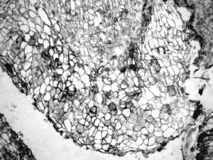
Section of a petiole of an unidentified plant infected with Synchytrium-like sporangia and zoospores
|
copyright: -
license: http://images.botany.org/index.html#license |
Image
|
Paleobotany

|

|
|
11-035h
|
|
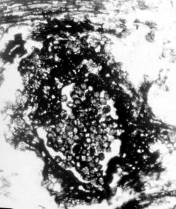
Cluster of Glomus-like chlamydospores occurring in enclosed structures, probable roots
|
copyright: -
license: http://images.botany.org/index.html#license |
Image
|
Paleobotany

|

|
|
11-036h
|
|
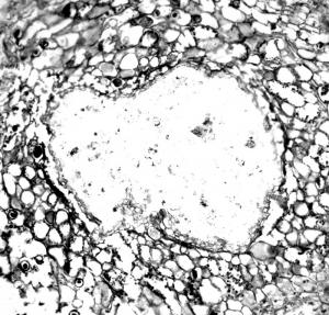
Section of a petiole of an unidentified plant infected with Synchytrium-like sporangia and zoospores
|
copyright: -
license: http://images.botany.org/index.html#license |
Image
|
Paleobotany

|

|
|
11-037
|
|
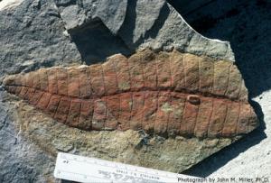
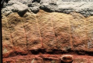
This is a kodachrome of a permineralized leaf fragment of Delnortea (USNM 387477). Taken by John M. Miller using a Nikkormat camera ASA 25 film in sunlight, shortly after collection of the rock sample from the Lower Permian (Leonardian) Road Canyon Formation, Permian Type Section. Later photographs were taken by Sergius H. Mamay, Ph.D., USNM and published as Fig. 15 in the American Journal of Botany 75(9): 1414.
|
copyright: none
license: - |
Image
|
Paleobotany

|

|
|
11-038
|
|
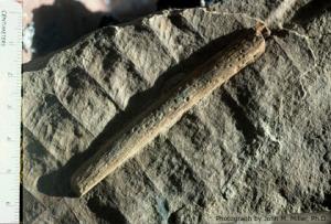
This is a kodachrome of a permineralized leaf fragment of the largest known fossil leaf of Delnortea (USNM 387473). Taken by John M. Miller, Ph.D. using a Nikkormat camera and ASA 25 film in sunlight, shortly after collection of the rock sample from the Lower Permian (Leonardian) Road Canyon Formation, Permian Type Section. Later photographs were taken by Sergius H. Mamay, Ph.D., USNM and published as Fig. 10 in American Journal of Botany 75(9): 1412.
|
copyright: none
license: - |
Image
|
Paleobotany

|

|
|
11-039
|
|
This is a kodachrome of a permineralized leaf fragment of Delnortea. The woody midrib, clearly visible on the slide, was used by William E. Stein, Jr., Ph.D. to create the polished thin-sections yielding the first images of a bifacial cambium and conducting xylem and phloem tissue of any Permian gigantopterid; shown as Figs. 23-36 (USNM 372427). Taken by John M. Miller, Ph.D. using a Nikkormat camera and ASA 25 film in sunlight, shortly after collection of the rock sample from the Lower Permian (Leonardian) Road Canyon Formation, Permian Type Section. Later photographs were taken by Sergius H. Mamay, Ph.D., USNM and William E. Stein, Jr., Ph.D. and published in AJB 75(9): 1418, 1420.
|
copyright: none
license: - |
Image
|
Paleobotany

|

|
|
12-001h
|
|
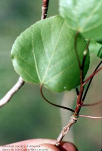
Populus tremuloides (quaking aspen) Laterally compressed petioles allow any breeze to cause leaves to vibrate or "quake". This movement makes it difficult for insects to land on or crawl on leaf surfaces.
|
copyright: John Curtis and Nels Lersten, BSA
license: http://images.botany.org/index.html#license |
Image
|
Plant Defense Mechanisms

|

|
|
12-002h
|
|
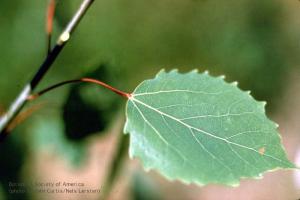
Populus grandidentata (big tooth aspen) Trichomes may form a dense mat that is a physical barrier to insects
|
copyright: John Curtis and Nels Lersten, BSA
license: http://images.botany.org/index.html#license |
Image
|
Plant Defense Mechanisms

|

|
|
12-003h
|
|
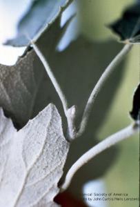
Populus alba with trichomes that remain on leaf.
|
copyright: John Curtis and Nels Lersten, BSA
license: http://images.botany.org/index.html#license |
Image
|
Plant Defense Mechanisms

|

|
|
12-004h
|
|
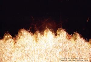
Populus grandidentata (young leaf) Dense mat of trichomes that abscise when leaf is fully expanded
|
copyright: John Curtis and Nels Lersten, BSA
license: http://images.botany.org/index.html#license |
Image
|
Plant Defense Mechanisms

|

|
|
12-005h
|
|
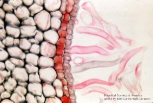
Populus grandidentata Young leaf with trichomes prior to abscission. Note constricted base of trichome - where abscission will occur.
|
copyright: John Curtis and Nels Lersten, BSA
license: http://images.botany.org/index.html#license |
Image
|
Plant Defense Mechanisms

|

|
|
12-006h
|
|
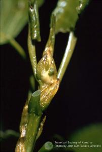
Populus deltoides Stem showing resin secreted by stipular bud scales.
|
copyright: John Curtis and Nels Lersten, BSA
license: http://images.botany.org/index.html#license |
Image
|
Plant Defense Mechanisms

|

|
|
12-007h
|
|
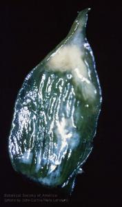
Populus deltoides Adaxial side of stipular bud scale with newly-secreted resin.
|
copyright: John Curtis and Nels Lersten, BSA
license: http://images.botany.org/index.html#license |
Image
|
Plant Defense Mechanisms

|

|
|
12-008h
|
|
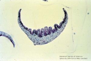
Populus deltoides Cross section of stipular buds scales - dark adaxial epidermis is composed of resin secretory cells.
|
copyright: John Curtis and Nels Lersten, BSA
license: http://images.botany.org/index.html#license |
Image
|
Plant Defense Mechanisms

|

|
|
12-009h
|
|
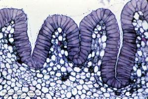
Populus deltoides higher magnification of adaxial surface of stipular bud scale. Dark cells are resin-secreting epidermis, light line above epidermis is cuticle pushed off by secretory product.
|
copyright: John Curtis and Nels Lersten, BSA
license: http://images.botany.org/index.html#license |
Image
|
Plant Defense Mechanisms

|

|
|
12-010h
|
|
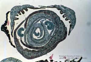
Populus deltoides bud cross section with stipular bud scales and young leaf lamina
|
copyright: John Curtis and Nels Lersten, BSA
license: http://images.botany.org/index.html#license |
Image
|
Plant Defense Mechanisms

|

|
|
12-011h
|
|
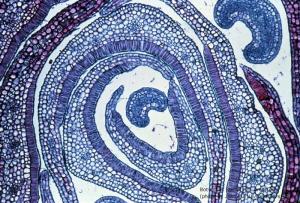
Populus deltoides higher magnification. Structures with dark stained adaxial epidermis is stipular bud scales.
|
copyright: John Curtis and Nels Lersten, BSA
license: http://images.botany.org/index.html#license |
Image
|
Plant Defense Mechanisms

|

|
|
12-012h
|
|
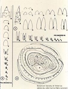
Populus deltoides bud anatomy (from Curtis & Lersten, Amer. J. Bot. 61:835-845. 1974) Fig. 1 represents dissected terminal bud (in cross section in Fig. 3). Fig. 2 represents dissected axillary bud.
|
copyright: John Curtis and Nels Lersten, BSA
license: http://images.botany.org/index.html#license |
Image
|
Plant Defense Mechanisms

|

|
|
12-013h
|
|
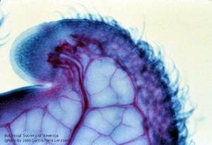
Populus deltoides leaf tooth clearing. Tooth tip is terminated by a resin gland.
|
copyright: John Curtis and Nels Lersten, BSA
license: http://images.botany.org/index.html#license |
Image
|
Plant Defense Mechanisms

|

|
|
12-014h
|
|
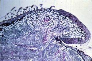
Populus deltoides 1 um paradermal section of leaf tooth showing resin secreting cells.
|
copyright: John Curtis and Nels Lersten, BSA
license: http://images.botany.org/index.html#license |
Image
|
Plant Defense Mechanisms

|

|